基礎生物学研究所


| Organizers | Jan Ellenberg (EMBL, Heidelberg), Naoto Ueno (NIBB) |
|---|---|
| Venue | Okazaki Conference Center, Okazaki, Japan |
| Date | Mar. 22-23, 2006 |
| Link | Symposium Website (http://www.nibb.ac.jp/emblsymp/2nd/) |
| Poster |
|
| March 22nd | |||
| Speaker | Affiliation | Time | Title |
|---|---|---|---|
| Registration | |||
| 8:40- | Registration | ||
| Opening Remarks | |||
|
|
9:05-9:15 | Opening Remarks | |
| Session1: Imaging diffusion & activity Chair: Yasushi Hiraoka | |||
| Atsushi Miyawaki | RIKEN, BRI, JAPAN | 9:15-9:50 | Visualization of the Spatial and Temporal Dynamics of Intracellular Signaling |
| Kai Johnsson | ISIC, EPFL, SWITZERLAND | 9:50-10:25 | Protein Chemistry in Living Cells |
| 10:25-10:50 | Coffee break | ||
| Tom K Kerppola | Univ. Michigan, USA | 10:50-11:25 | Visualization of Protein Interactions and Modifications in Living Cells using Bimolecular Fluorescence Complementation Analysis |
| Philipe Bastiaens | EMBL, GERMANY | 11:25-12:00 | Reaction-Diffusion Cycles in the Spatial Organization of Cellular Signaling and Morphogenesis |
| 12:00-13:00 | Lunch | ||
| Session2: Emerging technologies Chair: Peter T.C.So | |||
| Jennifer Lippincott-Schwartz | NIH, USA | 13:00-13:35 | Deciphering Protein Turnover, Topology and Transport in Living Cells |
| Masataka Kinjo |
|
13:35-14:10 | Analysis of Microenvironment of Nucleus Using Fluorescence Correlation Spectroscopy |
| Robert H. Singer | Yale Univ., USA | 14:10-14:45 | Following Single mRNAs in Living Cells |
| Akihiro Kusumi | Kyoto Univ., JAPAN | 14:45-15:20 | Single Molecule Tracking at the Cell Surface: Transient Signal Transduction by Engaged GPI-Anchored Receptors |
| 15:20-15:40 | Coffee break | ||
| Session3: Bioluminescence Chair: Tom K Kerppola | |||
| Susumu Terakawa | Hamamatsu Med. Univ., JAPAN | 15:40-16:15 | Exocytosis and Endocytosis in Neuronal Cells Visualized with Ultra High NA Lens |
| Tarik Issad | INSERM, FRANCE | 16:15-16:50 | The Use of BRET (Bioluminescence Resonance Energy Transfer) for the Study of Tyrosine-kinase Receptors |
| Takeaki Ozawa | IMS, JAPAN | 16:50-17:25 | Methods for Identifying Organelle-Targeting Proteins |
| Session4: Networks & Screening Chair: Philippe Bastiaens | |||
| Tobias Meyer | Stanford, USA | 17:25-18:00 | Fluorescence Microscopy Approaches for Dissecting Signaling Networks |
| Jan Ellenberg | EMBL, GERMANY | 18:00-18:35 | Microscopy-Based RNAi Screening and Quantitative Imaging of Chromosome Structure to Identify and Define the Function of Mitotic Genes |
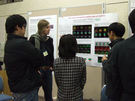
| 19:00-21:00 Mixer and Poster session |
|---|
| March 23rd | |||
| Speaker | Affiliation | Time | Title |
|---|---|---|---|
| Session5: 3D imaging Chair: Jan Ellenberg / Ernst H.K. Stelzer | |||
| Ernst H.K.Stelzer | EMBL, GERMANY | 9:00-9:35 | Selective Plane Illumination Microscopy: Life Sciences Require the Third Dimension |
| John W Sedat | UCSF, USA | 9:35-10:10 | OMX, A Microscope Platform for the Future? |
| 10:10-10:30 | Coffee break | ||
| Winfried Denk | MPI, GERMANY | 10:30-11:05 | Watching the Brain Compute and Tracing Its Wires: New Methods to Solve Old Riddles. |
| Haruo Kasai | NIPS, JAPAN | 11:05-11:40 | Dynamic Actin Organizations in Single Dendritic Spines of CA1 Pyramidal Neurons Studies with Two-Photon Photoactivation |
| Peter T.C. So | MIT, USA | 11:40-12:15 | Mechanotransduction: Understanding How Cells Sense Mechanical Signals with Novel Microscopic and Spectroscopic Tools |
| 12:15-13:00 | Lunch | ||
| Session6: Mitosis/Trafficking Chair: Atsushi Miyawaki | |||
| Jason Swedlow | Dandee Univ., UK | 13:00-13:35 | Studies of Mitosis in Living Cells and Tissues |
| Yasushi Hiraoka | NICT, JAPAN | 13:35-14:10 | Live-cell Observation of Chromosome Dynamics in Fission Yeast |
|
|
NIBB, JAPAN | 14:10-14:45 | Molecular Dissection of Membrane Dynamics during Autophagy |
| 14:45-15:05 | Coffee break | ||
| Session7: Organism model Chair: Jochen Wittbrodt / Naoto Ueno | |||
| Cornelis Weijer | Dandee Univ., UK | 15:05-15:40 | Chemotactic Cell Movement and Its Role in Morphogenesis |
| Damian Brunner | EMBL, GERMANY | 16:15-16:15 | Organizing Microtubules in Space and Time |
| Jochen Wittbrodt | EMBL, GERMANY | 16:15-16:50 | Individual Cell Migration as the Driving Force for Optic Vesicle Evagination |
| Kei Ito | Tokyo Univ. | 16:50-17:25 | Imaging the Structure of the Neural Circuits Using Molecular-Genetic Techniques |
| Minoru Tanaka | NIBB, JAPAN | 17:25-18:00 |
A Gonadal Field Coordinates Germline and Somatic Precursors to Form a Gonadal Primordium |

