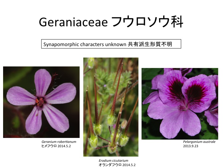
フウロソウ科のねじれた花柱 Twisted stigma of the Geraniaceae

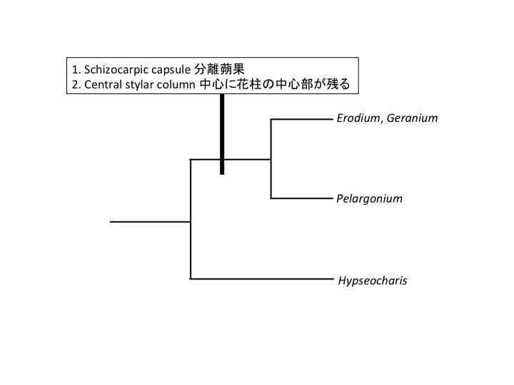
成熟時に個々の心皮が分離する分離蒴果。花柱中央部に集まった個々の心皮の辺縁部で個々の心皮が解離する。中央の軸は心皮が癒合することで中央に新しくできた組織だろう。花柱中心部と周辺部の間におそらく細胞壁の壊れやすい部分があり、この部分が壊れて、種子をつけた花柱表層がはじけ、種子散布を行う。

The middle layer of the stigma may contain cells with easily broken wall, where tissue dehisces for seed dispersal.

未成熟の果実では花柱基部は真っ直ぐだが、成熟した果実では花柱基部がよじれている。このよじれによって張力が貯まり、いきおいよく花柱が裂けて種子を散布する。どうしてこのようなよじれが生じるのだろうか。

A stigma is straight during the growth in seed maturation. When it is matured to dehisce, a stigma is twisted and produces power to disperse seeds.

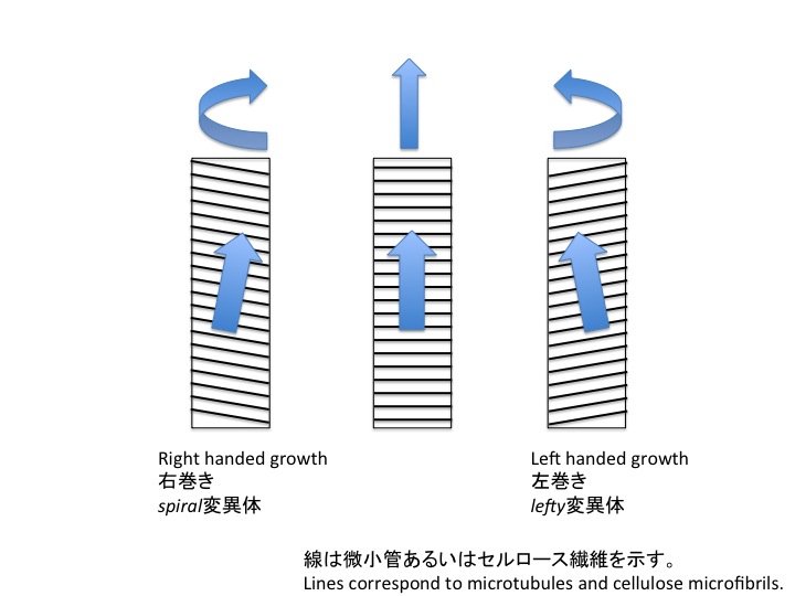
表層微小管は細胞壁のセルロース繊維の方向を制御すると考えられている(Griddings and Stachelin 1991)。セルロース繊維は細胞に巻き付いたたがのような役割をし、巻き方によって細胞の伸長方向が変わる。セルロース繊維が細胞の長軸に垂直な場合は細胞は長軸方向に伸長し(図中央)、このような細胞から構成されている器官は真っ直ぐ伸びる。一方、セルロース繊維が右肩下がりに配向すると、細胞は右上方向に伸長し(図左)、器官は右回りによじれる。逆に、左方下がりに配向すると(図右)、左回りによじれるようになる。TUBULIN遺伝子に突然変異のおきたspiral突然変異体、微小管結合タンパク質遺伝子に変異のおきたlefty変異体では、根、葉柄、花弁がそれぞれ右巻き、左巻きになる(Furutani et al. 2000; Thitamadee et al. 2002)。フウロソウの仲間の柱頭の果実形成時の成長過程で表層微小管の配向変化がおこり、よじれるのかもしれないが、研究例は無い。成長過程でどのようなシグナルによって表層微小管方向を変化させるのかもわかっていない。

The orientation of cortical microtubules likely regulate that of nascent cell wall microfibrils, which manages the direction of cell growth (Griddings and Stachelin 1991). Microtubules are oriented mostly perpendicular to the longer axis on a wild type Arabidopsis thaliana root cell, which elongates parallel to the longer axis (middle). When microtubules lean in right or left, the direction of cell elongation shifted and as a whole, a root composed of such cells is rotated (Furutani et al. 2000; Thitamadee et al. 2002). Similar mechanisms may function in Geranium pistil development.

Furutani, I. et al. 2000. The SPIRAL genes are required for directional control of cell elongation in Arabidopsis thaliana. Development 127: 4443-4453.

Griddings, T. H.Jr & Stachelin, L. A. in The Cytoskeletal Basis of Plant Growth and Form 85-100 (Academic, San Diego, 1991).

Thitamadee, S., Tuchihara, K. and Hashimoto, T. (2002) Microtubule basis for left-handed helical growth in Arabidopsis. Nature 417: 193-196.


カテゴリー: 5.14.04.01.Geraniales.フウロソウ目, Twisting ねじれ | フウロソウ科のねじれた花柱 Twisted stigma of the Geraniaceae はコメントを受け付けていません

コケシノブ科の包膜の多様性 Involucre diversity in the Hymenophyllaceae

Ebihara et al. (2006) A taxonomic revision of Hymenophyllaceae. Blumea 51: 221-280.

二弁状とカップ状になる形態は発生上のどのような違いによって生じているのだろうか。Iwatsuki (1977)は包膜の形成過程を観察し、発生過程によって異なった場所で細胞分裂が活発化するために二弁状になったりコップ状になることを示唆している。だとすると、時期場所特異的に細胞分裂を引き起こす機構がわかれば、二弁状包膜とコップ状包膜の作り方の違いがわかるのかもしれない。そもそも、組織のどこで細胞分裂を活発化させるかという問題は植物発生学、形態学全般に関わる問題であるがまだよくわかっていない。被子植物では葉の向軸型と背軸側の境界に分裂活性が生じることがわかり(Waites and Hudson 1995)、WOX遺伝子などが関与することがわかってきた(Nakata et al. 2012)。しかし、単葉の境界部分のどこがどのくらい伸長するかの制御機構はよくわかっていない。一方、複葉形成は葉原基の中で分裂活性の高いところと低いところを作ることによってできるが、その機構についてはオーキシン(Bilsborough et al. 2011)や転写因子(Vlad et al. 2014)の関与が明らかになってきている。この機構が単葉にもあてはまるかどうかは不明である。

しかし、そもそも、コケシノブの葉は1層の細胞しかないので、表裏をどのように決めているかは極めて不可解である。この点はセン類茎葉体についても同じである。セン類については葉の特定の部分で細胞分裂活性を制御する転写因子がありそうなので、今後研究を進めて、コケシノブにまで到達できると面白いと思っている。

How is the difference between bivalvate and cup-shaped involucres formed? Iwatsuki (1977) observed their development and showed that spatiotemporal difference of cell division activity caused the different morphology. When we know the molecular mechanisms to regulate such cell division patterns, we will get a part of the answer on the evolution of involucre divergence in the Hymenophyllaceae. Regulatory mechanisms of spatiotemporal cell division activity is fundamental question in developmental biology and morphology but we do not have clear answers. In angiosperms, the border between adaxial and abaxial domains have division activity (Waites and Hudson 1995) and WOX is involved (Nakata et al. 2012). However, mechanisms to regulate spatiotemporal activity regulation in the border is not well known. By the way, a compound leaf is formed differential division activity in a leaf primordium and the involvement of auxin (Bilsborough et al. 2011) and transcription factors (Vlad et al. 2014) have been reported.

Here we need to remember that a leaf of most filmy ferns is a single layer of cells. It is curious how they determine the adaxial and abaxial sides as well as the spatiotemporal division activity. This is similar in mosses. In the moss Physcomitrella patens, we found a transcription factor to regulate spatiotemporal cell division activity in a leaf and the study may be a breakthrough for understanding the leaf shape evolution and may be able to explain the diversity of involucres in the Hymenophyllaceae.

Bilsborough, G.D., Runions, A., Barkoulas, M., Jenkins, H.W., Hasson, A., Galinha, C., Laufs, P., Hay, A., Prusinkiewicz, P., Tsiantis, M. (2011) Model for the regulation of Arabidopsis thaliana leaf margin development. Proc. Natl. Acad. Sci. USA 108: 3424-3429.

Iwatsuki, K. (1977) Studies in the systematics of filmy ferns III. An observation on the involucres. Bot. Mag. Tokyo 90: 259-267.

Nakata, M., Matsumoto, N., Tsugeki, R., Rikirsch, E., Laux, T., Okada, K. (2012) Roles of the middle domain-specific WUSCHEL-RELATED HOMEOBOX genes in early development of leaves in Arabidopsis. Plant Cell 24: 519–535.

Vlad, D., Kierzkowski, D., Rast, M.I., Vuolo, F., Dello Ioio, R., Galinha, C., Gan, X., Hajheidari, M., Hay, A., Smith, R.S., et al. (2014). Leaf shape evolution through duplication, regulatory diversification, and loss of a homeobox gene. Science 343: 780-783.

Waites, R. and Hudson, A. (1995) phantastica: a gene required for dorsoventrality of leaves in Antirrhinum majus. Development 121: 2143-2154.

カテゴリー: 3.06.Hymenophyllales, 分裂場所制御, 未分類 | コケシノブ科の包膜の多様性 Involucre diversity in the Hymenophyllaceae はコメントを受け付けていません

アブラナ科、ヘリオフィラ節、ヘリオフィラ属Brassicaceae, Tribe Heliophileae, Heliophila

南アフリカに分布。属内形態多様性が大きい。Heliophila longifoliaは、花序の花と花の間が長く伸長する介在成長する総状花序intercalary racemesを形成し、蔓状になる。また、果実が成熟すると種子のできる部分がくびれる。どうして種子のできる部分が太くなって、種子の間の部分が細くなるのだろうか。アブラナ科にはシロイヌズナのようにほとんど太さがかわらない種類もある。胚珠から心皮に対して植物ホルモンのような液性情報、あるいは、圧力のような物理刺激が伝わってこのような発生様式を産み出しているのかもしれない。異なった組織間の作用による特殊形態の進化は、あちこちでおこっていそうだが、分子機構がほとんどわかっておらず、進化の機構も未解明である。

The genus Heliophila is distributed in South Africa. This genus is morphologically diversified. Heliophila longifolia has intercalary racemes, which elongate as a vine. The fruit morphology is like a string of beads. Interaction between ovules and surrounding carpel including phytohormones and pressure may function but details of the mechanisms and also the evolution have not been studied.

カテゴリー: 5.14.04.08.Brassicales.アブラナ目 | アブラナ科、ヘリオフィラ節、ヘリオフィラ属Brassicaceae, Tribe Heliophileae, Heliophila はコメントを受け付けていません

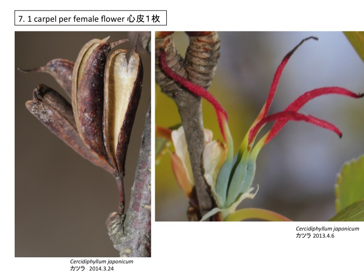
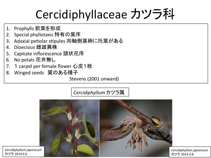
カツラ科 Cercidiphyllaceae

カツラ科は日本と中国に1属2種が分布。

Two species in one genus in a family are distributed in Japan and China.

prophyll 前葉


前葉は、イネ科など、側枝第一葉の形態が、その後に形成される葉と形態が異なることから名付けられた(Turpin 1819)。双子葉類と裸子植物では側枝の第1葉と第2葉(上図AからC)、単子葉類では側枝の第1葉(上図D)を指す場合が多いが、その後に作られる葉(上図の葉1や葉2など)と形態が異ならない場合が多い(熊澤1979)。双子葉類の場合は2枚の前葉は対生に蓋葉と主軸を結んだ線に直交するように側生し(上図AからC)、単子葉類では向軸側に1枚が生じる場合が多い(上図D)(熊澤 1979)。これら以外にも、いろいろな変異があり、多くの場合、融合や退化によって説明される(熊澤 1979)。双子葉類の場合、2枚の前葉が出た後、互生葉序になる場合(上図A、B)、背軸側(A)と向軸側(B)のそれぞれから次の葉が生じる場合がある。

Turpin, P.I.F. (1819). Mémoire sur l’inflorescence des Graminées et des Cyperées. Mem. Mus. d’Hist. Nat. Paris 5, 426–492.

熊澤正夫(1979)植物器官学、裳華房

Cercidiphyllum カツラ カツラの前葉は右図のように、太い葉脈が2本通っていることから、他の双子葉類に良く見られる側生する2枚の前葉が向軸側で癒合したものではないかと考えられている(熊澤1979)。一方、前葉は腋芽の蓋葉の托葉が癒合してできた可能性もある。あるいは、前葉は向軸側に1枚しかできず、その2枚の托葉が癒合したものである可能性もある(邑田仁博士私信)。カツラの前葉は2枚の前葉が癒合してできているとすると、前葉の葉腋には2個の腋芽が形成される可能性が高い。一方、托葉だとすると腋芽は形成されない。ただ、腋芽が退化消失している可能性もある。従って、2個の腋芽があれば、2枚の前葉が癒合した可能性が高くなるが、腋芽が無い場合は、2枚の前葉が癒合して腋芽が退化した可能性と、托葉が変形したという可能性の両方の可能性が残る。

Prophylls are the first leaves formed in the axillary shoot. A pair of opposite leaves are usually formed in dicots, while a single leaf in monocots. Morphological diversity of prophylls are explained by fusion or reduction. A prophyll of Cercidiphyllum japonicum has two main veins, suggesting the single prophyll is formed by a fusion of two prophylls (Kumazawa 1979). Another possibility is the fusion of two stipules of a subtending leaf (Dr. Jin Murata, personal communication) .
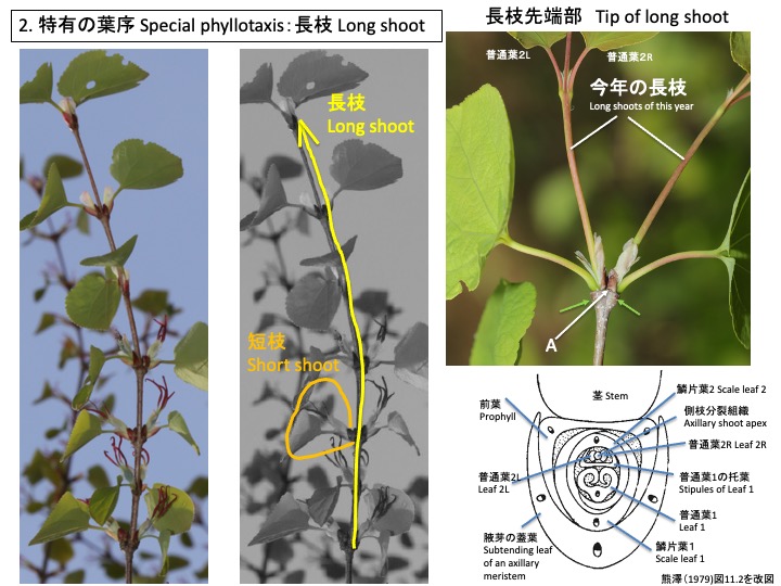
Cercidiphyllum japonicum カツラ カツラは、長枝と短枝を形成する。ともに仮軸分岐で、前年の葉の腋芽が伸長して新しい年の枝ができる。長枝の場合は、前年の茎頂が枯れ(図のA)、前年に一番先端で対生していた2枚の葉(図の黄緑矢印で落葉した後を示す)のそれぞれの腋芽が伸長する(図に「今年の長枝」と示す)。長枝先端の葉(蓋葉)の葉腋にできた側枝(腋芽)分裂組織は、前葉1枚、鱗片葉2枚、普通葉1枚を茎と蓋葉に直交するように2列互生する(注)。側枝分裂組織は、最初の普通葉(図の普通葉1)の托葉に包まれており、対生する普通葉を次々に形成する。図には、最初の1組の対生普通葉(普通葉2)を示す。最初は2列互生、後に対生に葉序を変化させる点で特有である(熊澤 1979)。

(注)長枝先端の腋芽を観察した報告が見つからなかったので、短枝の断面図(引用した熊澤1979図11.2)、長枝の先端を切ると先端よりの短枝が長枝に変わりうること(Titman and Wetmore 1955) から推定した。

Both long and short shoots of Cercidiphyllum japonicum are sympodial and a shoot of a next year is formed at the leaf axils of the previous year. A prophyll, two scale leaves, and a normal leaf are alternately formed in two rows in both types of shoots. In a long shoot, a lateral shoot meristem covered with stipules of the first normal leaf oppositely form subsequent normal leaves. The switch of alternate phyllotaxis to opposite one is special in Cercidiphyllum and some other angiosperms (Kumazawa 1979).

Cercidiphyllum japonicum カツラ 短枝も長枝と同様、蓋葉の葉腋にできた腋芽分裂組織は、前葉1枚、鱗片葉2枚、葉1枚を茎と蓋葉に直交するように2列互生する。長枝との違いは、普通葉1の托葉に包まれた側枝分裂組織が伸長せず、枯れてしまうか、花序となることである。従って、花序をつけない短枝には普通葉は1枚だけ形成される。他の樹木のように短枝に複数の葉を形成せず、1枚だけの葉を形成するため、数年を経た長枝でも、最初に長枝が成長した時のように主軸の両側に葉を対生して見える独特の枝ぶりとなる。

長枝の先端を切除すると直下の短枝は、短枝ではなく長枝を形成する。先端部を切除してもオーキシンを含んだラノリンペーストを塗布すると短枝の長枝化は阻害される(Titman and Wetmore 1955)。このことから、長枝の先端からのオーキシン供給によって下部の腋芽が短枝になるのではないかと推定されている(Titman and Wetmore 1955)。イチョウの短枝と長枝の区別にもオーキシンが関与していると推定されている(Gunckel and Wetmore 1946a, b, Gunckel and Thiman 1949, Gunckel et al. 1949)。シロイヌナズナではオーキシンに加えて、ストリゴラクトンが腋芽の休眠制御に関わっており、カツラやイチョウの場合も関与しているかもしれない。また、カツラは長枝と短枝で葉の枚数が異なっているが、イチョウの短枝は長枝とほぼ同じ数の葉を展開する。展開する葉の枚数を制御する仕組みはわかっていない。

The lateral shoot meristem of a short shoot has two fates. One is to be arrested and the other to form the inflorescence. However, the long shoot meristem is destroyed, lateral shoot meristems of short shoots change their fate to form a long shoot. The difference between long and short shoots should be regulated by auxin (Titman and Wetmore 1955). Similar auxin involvement between long and short shoot development is found in Ginkgo biloba (Gunckel and Wetmore 1946a, b, Gunckel and Thiman 1949, Gunckel et al. 1949). The same number of leaves are formed in a short shoot as that in a long shoot in Ginkgo biloba. Only one leaf is formed in Cercidiphyllum japonicum.

Gunckel, J.E. and Wetmore, R.H. (1946a) Studies of development in long shoots and short shoots of Ginkgo biloba L. I. The origin and pattern of development of the cortex, pith, and procambium. Amer. J. Bot. 33: 285-295.

Gunckel, J.E. and Wetmore, R.H. (1946b) Studies of development in long shoots and short shoots of Ginkgo biloba L. II. Phyl- lotaxis and the organization of the primary vascular system; primary phloem and primary xylem. Amer. J. Bot. 33: 532-543.

Gunckel, J.E. and Thimann, K.V. (1949) Studies of develop- ment in long shoots and short shoots of Ginkgo biloba L. III. Auxin production in shoot growth. Amer. J. Bot. 36: 145-151.

Gunckel, J.E., Thimann, K.V. and Wetmore, R.H. (1949) Studies of development in long shoots and short shoots of Ginkgo biloba L. IV. Growth habit, shoot expression and the mechanism of its control. Amer. J. Bot. 36: 309- 318.

Kumazawa, M. (1979) Plant organography (in Japanese). Shokabo

Titman, P.W. and Wetmore, R.H. (1955) The growth of long and short shoots in Cercidiphyllum. Amer. J. Bot. 42: 354-372.

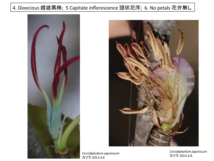
Cercidiphyllum japonicum カツラ


カテゴリー: 0.1.1.Shoot シュート, 5.14.01.Saxifragales.ユキノシタ目, Long and short shoot 長枝と短枝, Prophyll 前葉 | カツラ科 Cercidiphyllaceae はコメントを受け付けていません

カリッサ属 キョウチクトウ科 Carissa Apocynaceae

カリッサの棘は奇妙な位置についている。成長し終えた茎の先端にある棘を見ただけでは何が変形したのか首をかしげてしまう。しかし、「枝は必ず葉の腋に付く(芽は葉腋に形成される)」という被子植物の発生拘束を考慮すると仮説を考えることができる。

How are spines of Carissa formed? When we incorporate the developmental constraint of angiosperms: “new shoots are formed at the axils of leaves”, we can interpret that the spine is a shoot.

「枝は必ず葉の腋に付く(芽は葉腋に形成される)」ことを念頭に、まだ成長途中のシュート先端を観察すると、棘はシュートが変形したものだということがわかる。上の図で線は葉、矢印は茎を表してある。シュートは数対の葉を形成すると、腋に棘となるシュート(図の5と5’)を持つ小さな対生葉(2と2’)を作って茎頂分裂組織は成長を止める。棘となるシュートは対生葉をつけ、その腋にも棘となるシュートを形成する。

In the above drawings, lines and arrows indicate leaves and shoots, respectively. A main shoot forms several pairs of opposite leaves and terminates. The last two leaves (2 and 2’) are much smaller than previous leaves (e.g. 1 and 1’) and subtend a shoot to be a spine (5 and 5’).

シュート頂は棘となるか(左図)、さらに葉を着け、その腋にさらに棘となるシュートを形成する(右図)。

The spine shoot sometimes form leaves and the secondary spine shoots are formed at the axes of the leaves.


カテゴリー: 5.18.03.04.Gentianales.リンドウ目, Modified shoots | カリッサ属 キョウチクトウ科 Carissa Apocynaceae はコメントを受け付けていません

ビャクダン目の花被の起源 Origin of tepals in Santalales

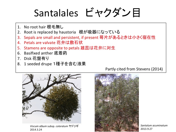
ビャクダン目は花の形態について、「1. Sepals are small and persistent, if present 萼片があるときは小さく宿在性」、「2. Petals are valvate 花弁は敷石状」、「3. Stamens are opposite to petals 雄蕊は花弁に対生」という共有派生形質を持つ。しかし、ビャクダン目の中には花被を1輪のみ形成し、花弁とガク片の区別ができないものがある。

Santalales has three synapomorphic characters on floral organs. However, some families have a single whorle of perianth instead of a calyx and a corolla.

Viscum album subsp. coloratum ヤドリギの花は雄蕊と対生する4枚の花被からなり2枚対生の苞葉(白矢印)を伴う。この花被は花弁、ガク片のどちらから進化したのだろうか。

A single whorle of four tepals opposite to stamens is formed in a flower of Viscum album subsp. coloratum of the Santaleceae. The flower is subtended by a pair of two bracts (white arrows). Which petals or sepals did evolve to the tepals?

Loranthaceae マツグミ科でもScurrula属グミバヤドリギ属の花も花被が1輪あるが、この花被が花冠と萼筒のどちらに起源したかは、わからない。

A single whole perianth is also formed in Scurrula of the Loranthaceae.

Loranthaceaeオオバヤドリギ科のLoranthus属の種は花冠の外側に小さな萼筒が形成される。このことから、Santalalesビャクダン目の花被は花弁と相同であると考えられる(Endress 1994)。被子植物全般において雄蕊と花弁は互生することが多いが、対生するものも見られる。互生、対生がどのような分子機構で制御されているのかは不明だが、被子植物全般の花形態進化に重要な問題であり面白い課題である。

Both a calyx and a corolla with five petals opposite to stamens are formed in Loranthus of the Loranthaceae, suggesting that tepals in Viscum and Scurrula are homologous to petals (Endress 1994).

Endress, P.K. 1994. Diversity and evolutionary biology of tropical flowers. Cambridge Univ. Press. Page 26.

Lindley, J. 1853. The vegetable kingdom: the structure, classification and uses of plants (3rd ed.). Bradbury and Evans, London.

カテゴリー: 5.15.Santalales.ビャクダン目, Perianths 花被 | ビャクダン目の花被の起源 Origin of tepals in Santalales はコメントを受け付けていません

Magnoliids モクレン類

カテゴリー: 5.05.01.Magnoliales.モクレン目, 5.05.02.Laurales.クスノキ目, 5.05.03.Canellales.カネラ目, 5.05.04.Piperales.コショウ目, Placentation 胎座, Spiral or Whorl 螺旋か輪か | Magnoliids モクレン類 はコメントを受け付けていません

アウストロベイレヤ目 Austrobaileyales

ANITAは基部被子植物の総称で側系統群。Amborellales, Nymphaeales, Illicidaceae, Trimeniaceae, Austrobaileyaceaeの頭文字をとったもの。真正双子葉類、マツモ目、単子葉類、モクレン類、センリョウ目の類縁はまだはっきりしていない。

ANITA group is paraphyletic and includes Amborellales, Nymphaeales, Illicidaceae, Trimeniaceae, and Austrobaileyaceae.

(1) Hardened mesotestal cells 外種皮中層の細胞が硬化、(2) Unilacunar nodes 単隙性葉脈分岐、の2つの形質が共有派生形質(Judd et al. 2008, Stevens 2001 onward) だが、肉眼レベルで観察できる共有派生形質は見つかっていない。

(1) Hardened mesotestal cells and (2) Unilacunar nodes are synapomorphic characters(Judd et al. 2008, Stevens 2001 onward) but no synapomorphic characters visible with naked eyes are not found.

アンボレラ目、スイレン目と同じように花被(花弁と萼片を併せた呼び方。両者を区別しにくいときに用いる)は、螺旋状に配列し、緑色の葉状の花被から色の付いた花弁状の花被へと順次に変化する。このことから、螺旋状に配列し、順次に変化する花被は、被子植物の原始形質(祖先が持っていた形質)である。

Austrobaileyales has spirally arranged tepals as Amborellales and Nymphaeales have, indicating this is a plesiomorphic character in angiosperms.

アウストロベイレア科は1科1属1種でオーストラリアの北部湿潤熱帯に自生し蔓性。「1. Flowers with internal staminodes 仮雄蕊が雄しべの雌しべ側にある」という形質が共有派生形質。

Austrobaileya scandens is a monotypic species in the Austrobaileyaceae, which is a liana and distributed in tropical Australia. “Flowers with internal staminodes” is synapomorphic character.

仮雄蕊は雌期には外側に開き、雌蕊が露出する。一方、雄期には雌蕊を多い雄蕊の葯が露出する。他花受粉促進のために仮雄蕊が機能している。

Staminodes open to expose a gynoecium in the female stage and close to cover a gynoecium in the male stage.

花被は外側から順番に形態を変える。ABCモデルは昆虫のホメオティック遺伝子による体節決定のように、ガク片、花弁、雄蕊、雌蕊が明確に器官分化している場合にはうまく適応できるが、器官形態が徐々に変化する場合にはうまく適用できない。また、シロイヌナズナのABCモデルのうちA機能については、他の被子植物で同じような機能を持っているかは不明であり、さらなる研究が必要である。徐々に器官形態を変化させる分子機構は植物独特かつ植物形態にしばしば見られるので、とても興味深い課題である。花被と雄蕊の境界は明瞭であり、ABCモデルでうまく説明できるように思われる。一方、仮雄蕊がどのような遺伝子によって制御されているのか、雄蕊と仮雄蕊、仮雄蕊と雌蕊の境界がどのように制御されているのかも不明である。

Staminode development as well as gradual morphological change in tepals are not well explained under the ABC model.

雄蕊は葉状になり、葯が向軸側に形成される。

Stamens are petaloid.

トリメニア科はパプアニューギニア、オーストラリア、フィジーに分布する。トリメニア属1属からなり、6種が知られる(Stevens 2001 onward) 。

Trimenia is monotypic in the Trimeniaceae, which includes 6 species and distributed in Papua New Guinea, Australia, and Fiji (Stevens 2001 onward) .

Trimenia属の葯は外向性、側向性の両方がある。時に内向性になることもある。Trimenia mooreiでは側向性である。茶色の花被は早落性。

Stamens are introrse, latrorse, or extrorse and vary between species. Stamens of Trimenia moorei is latrorse. Tepals in brown are caducous.

花被は外側のものはガク片的だが、ガク片と花弁の明瞭な区別はできない。花被は中心に近づくにつれてだんだん細くなる。

Morphology of tepals is gradually changed.

カテゴリー: 5.0.Angiosperms.被子植物, 5.03.Austrobaileyales.アウストロベイレア目 | アウストロベイレヤ目 Austrobaileyales はコメントを受け付けていません

カランコエの葉からの不定芽 Leaf plantlets in Kalanchoe

Kalanchoe tubifloraの葉は棒状で先端に複数(写真では4つ)の不定芽を形成する。この葉はどのような構造をしているのだろうか。

A petiole of Kalanchoe tubiflora is elongated and a leaf blade is reduced at the tip. Can you explain the curious morphology of the leaf blade?

葉身のでっぱった部分が葉の裂片でその間の凹部に不定胚が生じている。不定芽が大きくなるにつれて凹部がせり出し受け皿状になる。どうしてせり出すのかは不明。不定芽の影響があるのかもしれない。

Neelima Sinha研究室のGarcês et al. (2007)は、Kalanchoe daigremontianaの葉の不定芽発生過程を観察した。最初はドーム状に組織が隆起し、この形態はglobular stageの胚にも茎頂にも似ている。その後、ハート型胚に類似した形態になる。しかし、胚とは異なり、不定根が発生する。この不定根の発生様式はシュート下胚軸からの不定根形成に似ている。維管束は元の葉の維管束はと独立に形成される。これらのことから、不定芽は発生過程とシュートの両方のプログラムが混じって形成されているのではないかと推定されている。

Protruded regions are leaf lobes and plantlets are formed on sinuses. When plantlets become bigger, sinus tissue elongate to subtend a plantlet.

Garcês et al. (2007) described development of leaf plantlet of Kalanchoe daigremontiana. At first, tissue protrude as a dome and then becomes to be resemble to globular stage embryo as well as shoot apex. Then, the tissue becomes similar to a hart-shaped embryo. However, differently from an embryo, adventitious roots are formed. Vascular bundles are formed independent from leaf vasculature. The authors interpret that the development of leaf plantlet is a mixture of embryo and shoot development.

Garces, H.M., Champagne, C.E., Townsley, B.T., Park, S., Malho, R., Pedroso, M.C., Harada, J.J., and Sinha, N.R. (2007). Evolution of asexual reproduction in leaves of the genus Kalanchoe. Proc Natl Acad Sci U S A 104, 15578-15583.

カテゴリー: 5.14.01.Saxifragales.ユキノシタ目, Adventitious shoots 不定芽 | カランコエの葉からの不定芽 Leaf plantlets in Kalanchoe はコメントを受け付けていません

ホルトノキ科の蜜腺 Nectaries in the Elaeocarpaceae

ホルトノキ科のホルトノキ属Elaeocarpusは雄蕊の外側、花弁の内側に両者と互生の位置に蜜腺を形成する。一方、同科のTetrathecaPlatythecaは雄蕊外側に蜜腺を持たず、子房周縁から蜜を出している。Elaeocarpus species form extrastaminal nectaries. On the other hand, Tetratheca and Platytheca in the same family produce nectar from the surface of ovary.

ニュージーランドに広く分布するAristotelia serrataの雄花は、雄蕊外蜜腺と花の最中心部の蜜腺の2種類の蜜腺を持つ。観察した全ての雄花で雄蕊外蜜腺はあったが、花中央部蜜腺は未発達の雄花もあった。花最中心部蜜腺は雌しべの子房周縁の蜜腺が変化したものなのかもしれない。

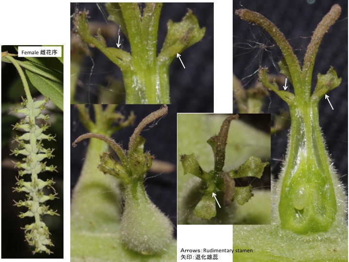
雌花は子房の周縁と、退化した雄蕊の外側に雄蕊外蜜腺が発達する。開花時には雄蕊外蜜腺は見えないが、果実が発達する段階で観察すると子房周縁の蜜腺の痕跡とともに、肉眼で見えるほど大きな雄蕊外蜜腺が形成されている。雌しべや雄しべが退化しても、雄蕊内蜜腺や雄蕊外蜜腺ができるということは、蜜腺が雌しべや雄しべに付随してできるのではなく、蜜腺形成機構として独立に存在している可能性を示唆しているのかもしれない。花器官の間にどのように蜜腺を形成するかの機構はまったくわかっていない。

A male flower of Aristotelia serrata have both extrastaminal nectaries and central nectary. The central nectary may be a modified extraovarial nectary. Only extraovarial nectaries are visible in a young female flower but both extraovarial and extrastaminal nectaries become conspicuous during fruit maturation.


カテゴリー: 02.Nectary, 5.14.03.03.Oxalidales.カタバミ目 | ホルトノキ科の蜜腺 Nectaries in the Elaeocarpaceae はコメントを受け付けていません