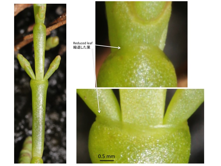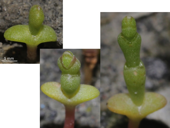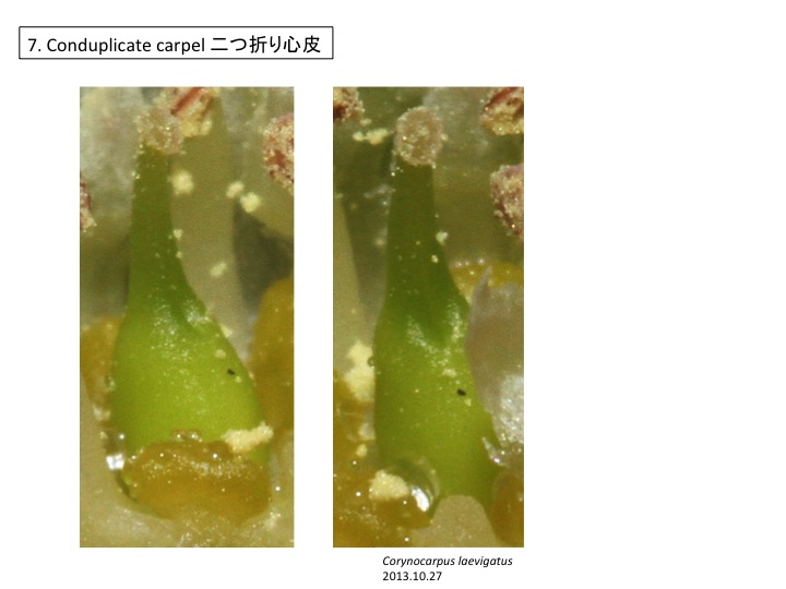
コリノカルプス科 Corynocarpaceae

Corynocarpus laevigatus, Corynocarpaceae

Corynocarpus laevigatus, Corynocarpaceae

コリノカルプス科はニューギニアから東オーストラリア、ニュージーランドにかけて分布する。

The Corynocarpaceae is distributed from New Guinea to Australia and New Zealand.

Corynocarpus laevigatus, Corynocarpaceae

Corynocarpus laevigatus, Corynocarpaceae

Corynocarpus laevigatus, Corynocarpaceae

Corynocarpus laevigatus, Corynocarpaceae

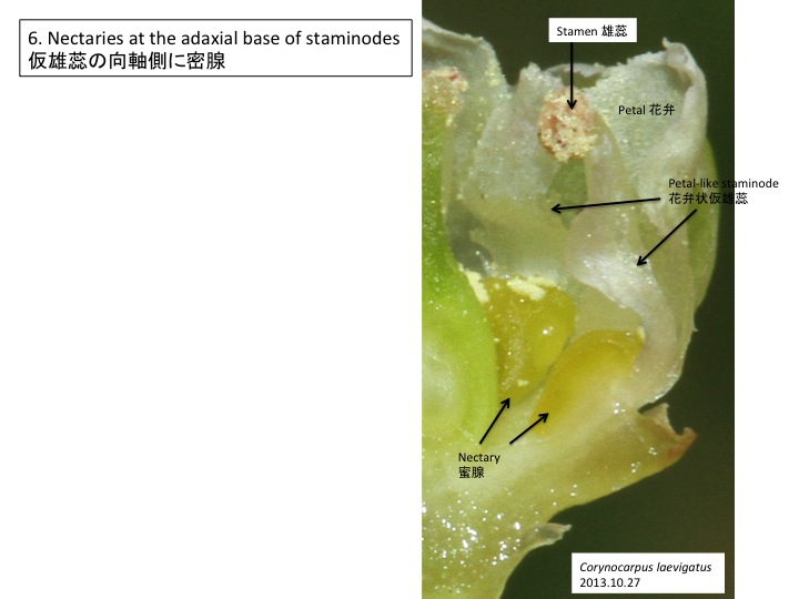
一部の雄蕊が花弁状に変化している。雄蕊で花弁形成遺伝子が異所的に発現すること、すなわちヘテロトピーによってこのような形態になっていると推定される。ヘテロトピーは形態多様性創出にしばしば関わっており、特に雄しべの花弁状仮雄蕊化は良く見られる。しかし、どうして異なった場所で適切に異なった発生過程の遺伝子が働きうるのかは不明。花器官形成遺伝子の発現場所を規定する因子の研究から道が開けるだろう。

Five stamens are petaloid, while five regular stamens are formed. The petaloid stamen, petaloid is likely formed by the ectopic expression of petal development genes, that is, heterotopy. Heterotopy is sometimes observed as a petaloid stamen also in other lineages. However, the molecular mechanisms how heterotopy occurs in flowering plants have not been reported.

Corynocarpus laevigatus, Corynocarpaceae

Corynocarpus laevigatus, Corynocarpaceae

カテゴリー: 5.14.03.07.Cucurbitales.ウリ目, Floral organ diversity in a flower 1つの花の中での花器官分化, 異所性異時性 heterotopy and heterochrony | コリノカルプス科 Corynocarpaceae はコメントを受け付けていません

サクライソウ目 Petrosaviales

Protosabiales

Petrosaviales サクライソウ目
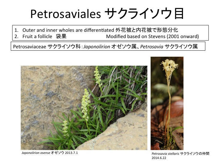
Petrosaviaceae サクライソウ科 Japonolirion オゼソウ属 Petrosavia サクライソウ属

Petrosaviaceae サクライソウ科 Japonolirion オゼソウ属 Petrosavia サクライソウ属

サクライソウ目にはサクライソウ科1科があり、日本固有のオゼソウ属と東アジアと東南アジアに分布するサクライソウ属の2属からなる。オゼソウは光合成能力のある葉を持ち独立栄養であるが、サクライソウ属は腐生である。サクライソウの仲間のPterosavia stellarisの葉緑体ゲノムが解読されており、光合成をしないためいくつかの遺伝子が欠損するとともにゲノムサイズが小さくなっている(Logacheva et al. 2014)。しかし、ATP合成酵素やrbcL遺伝子は残っており、光合成以外に必要なのかもしれない(Krause 2008)。rbcLについては脂質合成(Schwender et al. 2004)やRNA結合タンパク質(Yosef et al. 2004)として機能することが知られている。核ゲノムの解読によって、腐生化によって遺伝子レベルで何が変わっているのかが明らかになるだろう。

Pterosabiales contains a monotypic family Pterosaviaceae, which includes Japanolirion endemic to Japan and Pterosavia distributed in East and South Eastern Asia. Japanolirion osense forms green leaves and is autotrophic, while Pterosavia ssp. are saprophytic. Chloroplast genome of Pterosavia stellaris is reduced (Logacheva et al. 2014) but ATP synthase, rbcL and some other genes are still retained. The reason why these genes usually function in photosynthesis are retained is unknown but may function in other ways (Krause 2008). rbcL is revealed to function in lipid synthesis (Schwender et al. 2004) and as RNA binding protein (Yosef et al. 2004). No saprophyte genomes were published and such study will give insight on the evolution at the gene level.

Krause, K. (2008). From chloroplasts to “cryptic” plastids: evolution of plastid genomes in parasitic plants. Curr. Genet. 54: 111-121.

Logacheva, M.D., Schelkunov, M.I., Nuraliev, M.S., Samigullin, T.H., and Penin, A.A. (2014). The plastid genome of mycoheterotrophic monocot Petrosavia stellaris exhibits both gene losses and multiple rearrangements. Genome Biol. Evol. 6: 238-246.

Schwender J, GoVman F, Ohlrogge JB, Schachar-Hill Y (2004) Rubi- sco without the Calvin cycle improves the carbon eYciency of developing green seeds. Nature 432:779–782.

Yosef I, Irihimovitch V, Knopf JA, Cohen I, Orr-Dahan I, Nahum E, Keasar C, Shapira M (2004) RNA binding activity of the Ribu- lose-1, 5-bisphosphate carboxylase/oxygenase large subunit from Chlamydomonas reinhardtii. J Biol Chem 279:10148–10156.
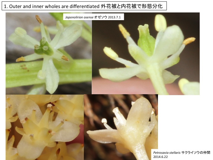
Petrosaviaceae サクライソウ科 Japonolirion オゼソウ属 Petrosavia サクライソウ属
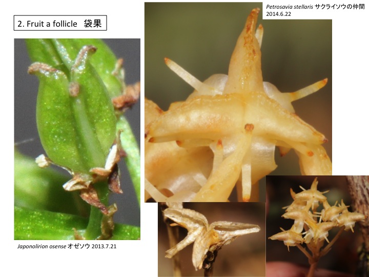
Petrosaviaceae サクライソウ科 Japonolirion オゼソウ属 Petrosavia サクライソウ属
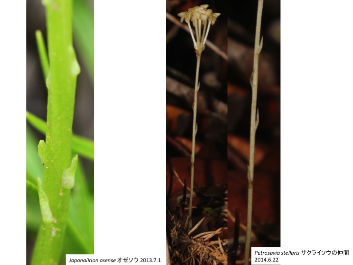
Petrosaviaceae サクライソウ科 Japonolirion オゼソウ属 Petrosavia サクライソウ属

Petrosaviaceae サクライソウ科 Japonolirion オゼソウ属

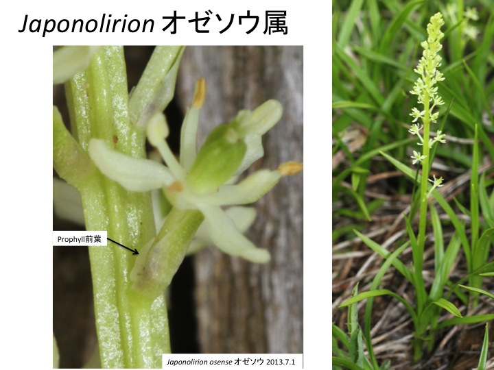
花茎上で各花に伸びる組織が花序茎表面に畝状に見える。各花に伸びる維管束が、花の形成される位置よりも基部側で花序維管束から分離しているのだろうか。花は苞と小花梗の向軸側に形成される前葉の2つの花器官によって抱かれている。他の分類群でも前葉は見られるが、どうしてあったりなかったりするのだろうか。

Ridges on an inflorescence stem are connected to flowers. It is unknown why this structure is formed in both structural and adaptive senses. A flower is subtended by a bract and a prophyll. The function of prophyll is unknown.

Petrosaviaceae サクライソウ科 Japonolirion オゼソウ属
Petrosaviaceae サクライソウ科 Japonolirion オゼソウ属 Petrosavia サクライソウ属

Petrosaviaceae サクライソウ科 Japonolirion オゼソウ属 Petrosavia サクライソウ属

オゼソウ、サクライソウの仲間ともに、花序の基部側に5個くらいの花を付けない苞が形成される。

In both Japanolirion and Pterosavia, bract-like organs are formed on the inflorescence.
 

カテゴリー: 5.06.03.Petrosaviales.サクライソウ目, mycoheterotrophic plant 菌従属栄養性植物, Prophyll 前葉 | サクライソウ目 Petrosaviales はコメントを受け付けていません

ブドウの蔓 Tendril of the Vitaceae

Vitis tendril

ブドウの蔓(Tendril)は葉と対生の位置に形成され、この形質はブドウ科の共有派生形質である。通常の植物では、葉と対生の位置に異なった器官ができることは無く、蔓の起源については多くの研究が行われてきた。一つの仮説は、蔓が主軸の先端で、葉の葉腋から腋芽が伸長し新たな主軸を形成する、即ち、仮軸分枝であるという仮説である。一方、主軸は単軸であり、蔓は一つ下の葉の腋芽で、それが茎に癒合したため葉と対生の位置に形成されるなど、他にもさまざまな仮説が提唱されてきた(reviewed in Shah and Dave 1970)。

しかし、切片を用いた細胞組織学的研究(Millington 1966; Tucker and Hoefert 1968; Sah 1960) から主茎頂から葉が形成されたあと、蔓が葉と対生の位置に新規に形成されることがわかった。このことから、ブドウ科の主茎は単軸であり、蔓は茎の側性の器官であると考えられている。しかし、どのような発生機構の変化によってこのような他の植物には見られない体制が進化したのかは不明である。また、蔓だけでなく蔓とシュートを併せ持った器官を形成する場合もあり、いつ幹細胞が形成されるのだろうか。蔓が形成初期から幹細胞を伴って形成されるのか、あるいは、葉と同じように幹細胞は無く、シュートを伴う場合には発生後期に幹細胞が形成されるのかといった問題は、幹細胞マーカー遺伝子の発現や葉序を制御するオーキシンが茎頂付近でどのような分布をしているかを調べることで解決できるかもしれない。

Tendrils of Vitaceae species are formed oppositely to leaves. Several hypotheses have been proposed (reviewed in Shah and Dave 1970). One hypothesis is that a tendril is a main shoot tip and a main shoot in appearance is an lateral shoot formed at a leaf axil. Other hypotheses include a hypothesis that a shoot is monopodial and a tendril is axillary formed and fused with a stem until the opposite position of an upper leaf.

However, Cytohistological studies showed that a tendril primordium is formed after the initiation of its opposite leaf, during which a shoot meristem of a main stem is maintained (Millington 1966; Tucker and Hoefert 1968; Sah 1960). This indicates that a main shoot is monopodial and tendrils are lateral organs. A reason why such lateral organs are formed at the opposite positions to leaves is still unknown. Tendrils sometimes accompany a shoot (see a right bottom picture) but it is unknown when the new shoot meristem is initiated.

Millington, W.F. 1966. The tendril of Parthenocissus insert: Determination and dvelopment. Amer. J. Bot. 53: 74-81.

Sah, J.J. 1960. Morphohistogenic studies in Vitaceae. I. Origin and development of the axillary buds in Cayratia carnosa Gagnep. Phytomorphology 10: 157-174.

Shah, J.J. and Dave, Y.S.(1970) Morpho-histogenetic studies on Tendrils of Vitaceae. Amer. J. Bot. 57: 363-373.

Tucker, S.C. and Hoefert, L.L. 1968. Ontogeny of the tendril in Vitis viniferra. Amer. J. Bot. 55: 1110-196.

カテゴリー: 5.14.02.Vitales.ブドウ目, Tendril | ブドウの蔓 Tendril of the Vitaceae はコメントを受け付けていません

アワブキ目 Sabiales

アワブキ目はアワブキ科1科からなり、東南アジア、中南米の熱帯域を中心に分布。

The Sabiales is composed of a single family the Sabiaceae, which is mainly distributed in tropics of Southeast Asia and Central and South America.

<Meliosma arnottiana subsp. oldhamii フシノハアワブキ; Meliosma myriantha アワブキ; Sabia japonica アオカズラ>

アワブキ属は25から70種からなり、東南アジアと中南米の熱帯域に分布する (Ronse De Craene and Wanntorp 2008)。

Meliosma includes approximately 25 to 70 species and is distributed in tropical areas of Southeast Asia and Central and South America (Ronse De Craene and Wanntorp 2008).

Ronse De Craene, L. P. and Wanntorp, L. 2008. Morphology and anatomy of the flower of Meliosma (Sabiaceae): Implications for pollination biology. Plant Syst. Evol. 271: 79 – 91.

<Moliosma cuneifolia; Meliosma myriantha アワブキ; Meliosma arnottiana subsp. Oldhamii フシノハアワブキ>

アワブキの花はらせん配列した5枚のガク片(図3:花を花梗側から写してある)、3枚の大型の花弁と2枚の細い花弁(図4の黒星)、1室からなる半葯を2つ持つ雄蕊が2本(図4の赤星)、3枚の仮雄蕊(図4の青星、図5に各仮雄蕊の境界を灰色の線で示す)を形成する。2枚の心皮が癒合した雌蕊の柱頭は3枚の仮雄蕊の間から飛び出す(図1の矢印)。仮雄蕊には葯が入るように窪みができている(図2;図5にへこみの位置を破線で示す)。窪みの中でできた葯は雄蕊が成熟し葯糸が背軸側にそると(図6)、細い花弁にぶつかって、花弁上に花粉をまき散らし、花粉散布に役立つ(Ronse De Craene and Wanntorp 2008)。窪みは葯がぴったりはまる大きさにできているが、どのような仕組みでできるのだろうか。二つの器官の相互作用によって形態ができる仕組みはまだよくわかっていない。また、5枚の花弁のうちどうやって2枚の花弁の形を変化させる仕組み、そして、2本の雄蕊と3本の仮雄蕊を作り分ける仕組みも不思議だ。

A flower of Meliosma myriantha is composed of spirally arranged five sepals (Fig. 3), three large and two thin petals (Black asterisks in Fig. 4), two stamens with two thecae with a locule (red asterisks in Fig. 4), three staminodes (blue asterisks in Fig. 4; borders of three staminodes are indicated by gray lines), and a gynoecium whose pistil is protruded among staminodes (an arrow in Fig. 1). When stamens are matured, filaments are inverted outward and anthers hit against thin petals, which is effective for pollen presentation (Ronse De Craene and Wanntrop 2008). Hollows in staminodes fit anthers. The mechanism how to adjust the size, that is morphogenesis caused by interaction between two organs, is unknown. It is also interesting to know the mechanisms to make different types of petals as well as stamens and staminodes.

Ronse De Craene, L. P. and Wanntorp, L. 2008. Morphology and anatomy of the flower of Meliosma (Sabiaceae): Implications for pollination biology. Plant Syst. Evol. 271: 79 – 91.

<Meliosma myriantha アワブキ >

カテゴリー: 5.09.01.Sabiales.アワブキ目, Floral organ diversity in a flower 1つの花の中での花器官分化, Leaf vein 葉脈, Pressure and morphogenesis | アワブキ目 Sabiales はコメントを受け付けていません

ヤマモガシ目とアワブキ目の姉妹関係の共有派生形質 Synapomorphic character to connect Proteales and Sabiales

アワブキ科はガク片、花弁、雄蕊がすべて互いに対生の位置にある。ヤマモガシ科は1輪の花被を持ち、花被片が雄蕊と対生している。被子植物では、ガク片、花弁、雄蕊が互いに互生の位置関係にあることがもっとも多いので、対生になるのは派生的形質である。アワブキ属の種の研究から、各器官原基は螺旋状に形成されるが、そのタイミングがずれるために対生になることが知られている(Wanntorp and Ronse De Craene, 2007)。真正双子葉類では、螺旋配列する花器官原基のできるタイミング、すなわち葉間期plastochronが短くなることによって輪になる(Endress 2011)。花器官の多様性を導く機構として、各々の花器官がどのようなタイミングで出来るか、即ち、葉間期の長さ、あるいは、葉原基成長のタイミング、の制御機構がわかると、花器官の多様性の多くの部分が説明できるようになると思う。

The Sabiales includes the Sabiaceae and the Proteaceae. Sepals, petals, and stamens are oppositely arranged each other in the Sabiaceae. In Proteaceae, each tepal is opposite to each stamen. Most eudicots have alternative arrangement and the opposite arrangement is synapomorphic in the Sabiales and Proteales. Floral organs are spirally formed and the opposite arrangement is caused by the change of plastochron length in Meliosma in the Sabiaceae (Wanntorp and Ronse De Craene 2007). A whole structure of floral organs in eudicots is formed because the length of plastochron is shorten (Endress 2011). If we know the molecular mechanisms to regulate plastochron length, we will be able to understand how diversity of floral organ arrangement evolved.

Endress (2011) Evolutionary diversification of the flowers in angiosperms. Amer. J. Bot. 98: 370-396.

Wanntorp L., Ronse De Craene L.P. (2007) Flower development of Meliosma (Sabiaceae) – evidence for multiple origins of pentamery in the eudicots. Amer. J. Bot. 94: 1828–1836.

<Sabiales アワブキ目; Sabiaceae アワブキ科; Sabia japonica アオカズラ; Meliosma myriantha アワブキ; Proteales ヤマモガシ目 Proteaceae ヤマモガシ科: Isopogon formosus>

カテゴリー: 5.09.01.Sabiales.アワブキ目, 5.09.02.Proteales.ヤマモガシ目, Plastochron 葉間期, Spiral or Whorl 螺旋か輪か | ヤマモガシ目とアワブキ目の姉妹関係の共有派生形質 Synapomorphic character to connect Proteales and Sabiales はコメントを受け付けていません

仮軸分枝と葉序 Sympodial branching and phyllotaxis in Stellaria

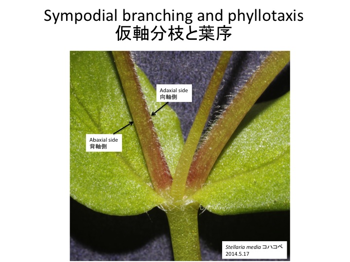
コハコベは茎の片側に毛を叢生する。毛の生える方向は決まっていて、葉腋から腋芽が伸びるときに、主軸側(向軸側)に毛を叢生し、背軸側にはほとんど毛を生じない。

花は茎と連続しその頂端に形成されるので、茎と同じように向軸側に毛を叢生する。左の写真の花梗は背軸側が見えているので毛がほとんど無い。右の図は花と花梗を横から写したものである。毛の生えている側が向軸側である。

In Stellaria media, hair is formed on one side of a stem. When two shoots are formed from axils of opposite leaves, hair is formed on the adaxial side of each shoot. 

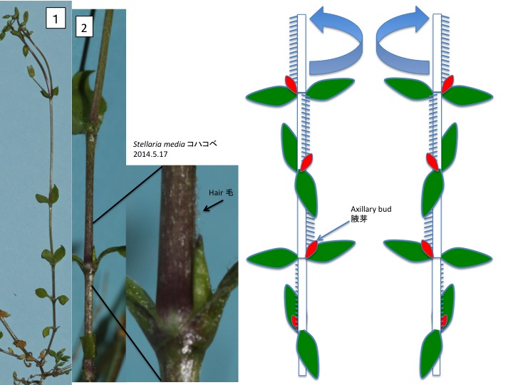
コハコベの栄養シュートは写真1のように、一見すると1本の茎が十字対生葉序を付けてまっすぐ伸びている単軸のように見える。ところがどうもそうではないようである。コハコベの栄養シュートの茎を見ると前のページの集散花序の腋芽から伸びた茎のように片側に毛が生えている。ところが、毛は節間茎(葉と葉の間の茎)ごとに生える方向が変わっていて、90度ずつずれている。どうしてだろうか。腋芽の付く位置を見ると、写真2のようにいつも毛と向かい合う位置に付いている。ここで前のページの葉腋から2本の腋芽が出る場合を思い出して欲しい。左右それぞれの葉の腋芽由来のシュートは向軸側を向くように毛が生えていた。いつも右か左のどちらか同じ側の腋芽だけが伸びて他方の腋芽が伸びないとすると、栄養シュートの茎の毛の付き方と同じパターンになる。葉は十字対生していて、90度ずつずれて回転しているので、毛の向きが90度ずつずれるのは葉序の回転と対応している。このことから、コハコベの栄養シュートの茎は仮軸分枝によってできていると考えられる。

Linearly aligned hair is formed in one face of a stem in Stellaria media and the direction rotates 90 degree. When we observe the node, an axillary bud is always faces to hair. Shall we remember that hair is formed on the adaxial side of axillary shoots in the cyme, which is shown in the previous page. It is possible to interpret that a vegetative shoot of Stellaria media is formed by sympodial branching, although it looks like a monopodial shoot. The rotated direction varies among stems.

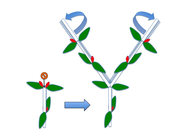
コハコベの茎が分岐するときは、2枚の対生葉の間にあった茎頂分裂組織が消失し、かわりに、両方の葉腋の腋芽が伸び出す。まだ、数個対観察しただけなので確信は持てないが、どうも、左右のシュートで毛の回転方向、すなわち、葉序の回転方向が逆になっているようである。葉序はオーキシンの移動を考えることでかなり説明できるようになってきた(総説としてTraas 2013)。コハコベに見られるような葉序の転換はどのように基本システムを変えるとできるのだろうか。

維管束植物の祖先の茎はもともと二叉分枝で、原生の小葉類や一部のシダ類にそのなごりが見られる。二叉分岐からどのように発生過程を変化させることによって単軸の茎が進化してきたのかはよくわかっていない。茎の分岐様式の進化は陸上植物の体制進化において大きなイベントの一つであり、主軸形成機構の進化がそれを担っていた可能性がある。主軸形成機構とその進化に関するヒントが被子植物における仮軸分枝の研究から得られるかもしれない。

When two axillary shoots are formed at the node, they have different rotated directions. Phyllotaxis is well explained by the auxin movement (reviewed in Traas 2013). However, it appears that it is still difficult to explain this type of switching..

One of conspicuous innovation of body plans in land plants is the change from dichotomous branching to monopodial branching but the evolution of the development of branching is mostly unknown. Developmental and molecular mechanisms of overtopping appears one of keys to solve the problem and detailed studies of overtopping in sympodial branching may give insight.

Traas, J. (2013) Phyllotaxis. Development 140: 249-253.

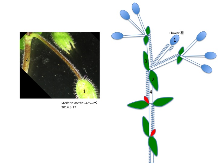
栄養茎から集散花序に転換するとき、茎頂は花(図の1の花)で終わる。花梗の毛は直前の茎(図の4)の毛の向きと同じである。葉腋からシュートが形成されるときは毛の向きが変わるが、頂端に付く花は茎の延長なので葉序が変わらないのだろう。

栄養茎の最先端の2枚の葉(図の2と3)の腋からそれぞれ回転方向の違う二次花序が伸長する。上図では二次花序の葉腋から花が出て終わっているので二次花序の葉序の巻き方向はわからないが、二次花序の葉腋にさらに三次花序を形成することもあり、その際には葉序の巻き方が互いに逆になる。

花序の末端付近に付く花の花梗では毛がみられなくなる。

A shoot is terminated with a flower (1 in Figure). The direction of terminal flower pedicel hair is the same as the connected stem (4 in Figure). When new shoots are formed at the axils of two leaves (2 and 3 in Figure), two shoots twist in different directions each other.

カテゴリー: 0.1.1.Shoot シュート, 5.17.Caryophyllales.ナデシコ目, Phyllotaxis 葉序, Sympodial branching 仮軸分枝 | 仮軸分枝と葉序 Sympodial branching and phyllotaxis in Stellaria はコメントを受け付けていません

ハコベ属の集散花序 Cyme in Stellaria

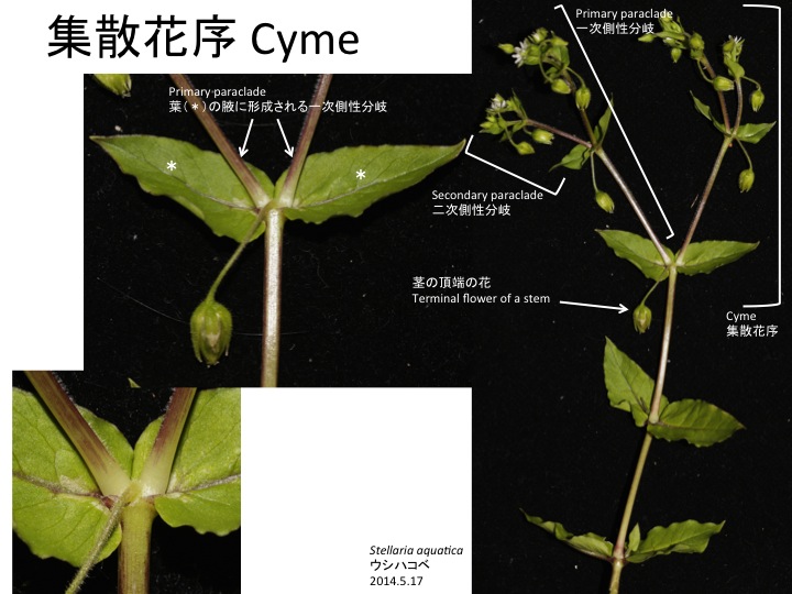
ウシハコベの集散花序では、まず茎頂が花原基に変化する。そして、茎頂に近い葉の腋から側性分岐(一次側性分岐)が生じ、1対の葉を付けた後、その茎頂が花原基に変化する。すると、一対の葉の腋から新たな側性分岐(二次側性分岐)が生じ、同じようなパターンを繰り返し、集散花序を形成する。

A shoot of Stellaria aquatic terminates with a flower and primary paraclades are formed at the axils of uppermost leaves. Then the 1st paraclades form a pair of leaves and terminate with a flower. Secondary paraclades are formed at the axils of the first paraclade leaves. As a result, a cyme is formed.

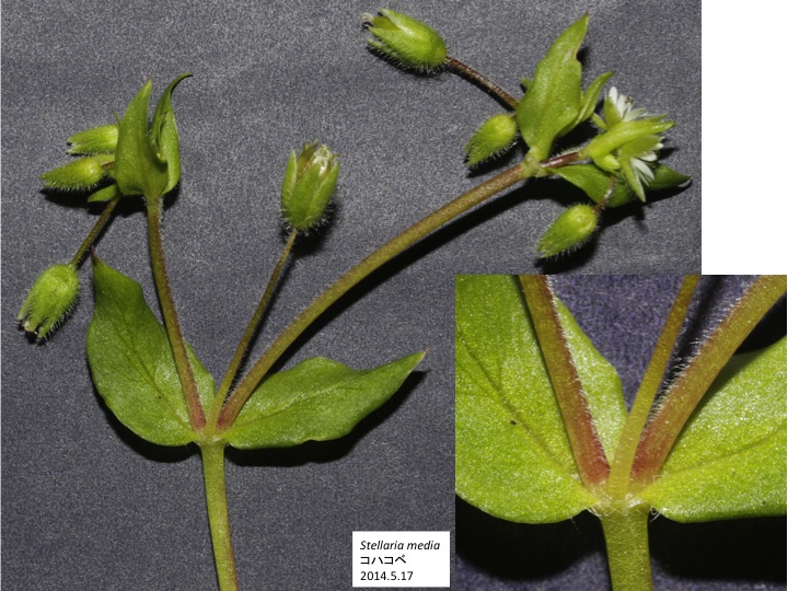
コハコベもウシハコベと同じように集散花序を形成する。

Stellaria mudia forms a cyme as S. aquatica.

カテゴリー: 5.17.Caryophyllales.ナデシコ目, Inflorescence 花序, Phyllotaxis 葉序, Sympodial branching 仮軸分枝 | ハコベ属の集散花序 Cyme in Stellaria はコメントを受け付けていません

Sinowilsonia シノウイルソニア属

Sinowilsonia属はユキノシタ目マンサク科マンサク亜科Eustigmateae節。中国に固有。

The genus Sinowilsonia belongs to Eustigmateae, Hamamelidoideae, Hamamelidaceae, Saxifragales and is distributed in China.

雄、雌、両性の花序を形成する。

Male, female, and hermaphrodite inflorescences are formed in a tree.

両性花序の基部10個ほどは両性花でそれ以外の花は雄花。雄花序の全ての花は雄花。雄花は扁平化して花被と雄蕊が癒合した雄蕊花被複合体を形成する奇妙な形態。クルミ科でも雄花序が扁平化し花も扁平化している。陸上植物の形態進化仮説であるテローム説 (Zimmermann 1953, 1965) では立体的な分岐した茎が扁平化して葉になったという仮定をおくが、Sinowilsoniaでおきた扁平化の仕組みを知ることが出来れば、テローム説についてさらに踏み込んだ検討ができるかもしれない。

Basal 10 flowers are hermaphrodite in a hermaphrodite inflorescence and other flowers are male. All flowers are male in male inflorescences. Each male flower is flattened and each tepal is fused with each stamen in an opposite position. Male inflorescences and flowers of Juglans  are flattened and may use similar mechanisms in Sinowilsonia. The mechanism of the flattening may give insight to the “Planation” of the Telome theory (Zimmermann 1953, 1965).

Zimmermann, W. 1953. Main results of the “telome theory.” The Palaeobotanist 1: 456-470.

Zimmermann, W. 1965. Die Telomtheorie. Gustav Fischer Verlag, Stuttgart.

雄花序先端部のいくつかの扁平化した雄花。

Flattened male flowers in a distal part of a male inflorescence.

両性花序基部に10個ほど両性花を形成する。雄花同様、雄蕊とそれと対生の位置にある花被が癒合し、雄蕊花被複合体を形成している。さらに、5本の雄蕊花被複合体が癒合し筒状になっている。さらに、その外側に白い毛が生えた器官が癒合しているように見える。この器官は何であろうか。花被、小苞、苞の可能性があるがどれかは発生過程、発現する遺伝子を調べればわかると思う。

Approximately 10 flowers are formed at the base of a hermaphrodite inflorescence. Five stamen-tepal complexes are fused to cover gynoecium. Furthermore, a layer of tissue with white hair covers. It is unknown the white tissue is an outer tepal, bracteole, or a bract until we investigate the developmental process and expression patterns of genes involved in these organ development.

雌花序は雌花のみから構成されている。雌花は両性花のように、雌蕊を雄蕊花被複合体が覆い、その外側を白い毛の生えた器官が覆っている。雌花の雄蕊花被複合体は葯が無く、短い葯糸のみがある。さらに、花糸が花被と癒合せず、花被は両性花や雄花の花被に較べて大きく、切れこんでいる。葯と花被の大きさはトレードオフの関係にあるのだろうか。

A female inflorescence is composed of female flowers. In a female flower, a gynoecium is covered with a fused stamen-tepal complexes and a fused organ covered with white hair as in a hermaphrodite flower.  However, anthers are rudimental and shorter filaments than those in hermaphrodite flowers are not fused with tepals. Tepals are bigger and more lobed than those in hermaphrodite flowers.


カテゴリー: 5.14.01.Saxifragales.ユキノシタ目, Telome | 2件のコメント

Schizaeales フサシダ目

フサシダ目においてどうして環帯は胞子嚢の先端部にできるのだろうか。シダ類の他の群でも環帯の位置は目レベルで保存されている場合がほとんどである。一見、単純な構造のようにも思えるが、環帯は、胞子嚢を裂開し胞子を飛散させる働きをしており、機能としては複雑である。環帯の形が少しでも変わると胞子がうまく飛散できなくなってしまうので、環帯の位置は変わりにくいのかもしれない。しかし、では、どうやって、現在の環帯はできたのだろうか。シダ類の祖先から変化してこのような形に落ち着いたと考えられるが、変化の過程では胞子散布が非効率になるなどのデメリットはなかったのだろうか。いろいろな可能性は考えられるが、まずは、環帯がどのような分子機構によって形成され、異なった形態の環帯はなにがどうかわることによってできるのかの詳細を明らかにしてから議論をすることが必要だと思う。

Why is the position of annulus conserved in the Schizaeales? Even in other groups of ferns, the position of annulus is well conserved in a family or order. Annuli look simple at a glance but the function to open a sporangium and disperse spore should be nicely adjusted. So it may be difficult to accumulate mutations causing slight change to the annulus position, which makes the mutants to be less adaptive than their parents. The annulus position of the Schizaeales may be at the top of local adaptive optimum and may not be shifted. Anyway, the molecular mechanism to regulate the annulus positions are not known at all and future study will give insight on the evolution of the position.

カニクサ Lygodium japonicumの根には主根と異なった色合いをした菌根が形成される。菌根の内側にはアーバスキュラー菌根菌が共生する大きな細胞が並んでいる。アーバスキュラー菌根菌と共生することでリンの取り込みが増え、成長が良くなることが調べられている(Soti et al. 2014)。菌根菌との共生は陸上植物進化の初期から起こっており、多くのシダ植物でアーバスキュラー菌根菌との共生が報告されている(Wang and Qiu 2006)。

Many mycorrhiza by arbascular mycorrhizal fungi were formed on roots of  Lygodium japonica. Higher phosphorus uptake and better growth with the symbiosis is reported in L. microphyllum (Soti et al. 2014). Symbiosis between plants and arbascular mycorrhizal fungi was originated at the early stage of land plant evolution and many pteridophytes form mycorrhiza.

Soti, P.G., Jayachandran, K., Purcell, M., Volin, J.C., and Kitajima, K. 2014. Mycorrhizal symbiosis and Lygodium microphyllum invasion in South Florida – a biogeographic comparison. Symbiosis 62: 81-90.

Wang, B. and Qiu, Y.-L. 2006. Phylogenetic distribution and evolution of mycorrhiza in land plants. Mycorrhiza 16: 299-363.

一対の胞子を形成する羽片が立ち上がり、立体的な葉を形成する。一部の羽片だけに胞子嚢を形成する種はシダ類の中にしばしば見られるが、胞子嚢を形成する羽片が立体的になる種はAnemia属以外には無く、どうしてこのような複雑な形態を作れるのかの仕組みは面白いと思う。

A pair of basal pinna becomes fertile and bears sporangia. It is often observed in ferns that a part of pinna become fertile but quite unique that fertile pinna become upward. The mechanism to form such more complex leaf shape is still unknown.

フサシダ科の葉は葉柄と葉身の区別がつかず、葉の先端にいくつかの稔性裂片、すなわち胞子嚢を形成する裂片を形成する。

A petiole and a lamina are not distinguishable in leaves of the Schizaeaceae. Several fertile pinna bearing sporangia are formed at the tips of a leaf.

カテゴリー: 3.08.Schizaeales | Schizaeales フサシダ目 はコメントを受け付けていません

Swartzieae イチベンバナ連

Swartziaイチベンバナ属の花は雄蕊が多数あり、2形成を示す。ほとんどの雄蕊は花弁の側(向軸側)に立ち上がるが、何本かが他の雄蕊よりも大きく反対側に伸長する。Swartzia pinnataでは2本の太くて大きな葯を付けた雄蕊が雌しべの方に下降する。花原基で雌蕊原基の周りを覆うようにリング状の分裂組織(ring meristem)ができ、そこから大きな雄蕊になる原基が膨らみ、残った向軸側部分からたくさんのより小さな雄蕊原基が形成される(Tucker 2003)。どうやって異なった形態の雄蕊を作るのか、両者の位置をどのように決めているのかは未解明。

1つの花の中に異なった形態の雄蕊を持つことをheteroanthery異型雄蕊性と呼ぶ (Endress 1994)。Swartziaの場合、花弁の手間にあるのがハチに食用として花粉を提供する雄蕊 feeding stamensで、それらの下側により大型で隠れたように本来の目的である受粉用の雄蕊がある。ハチによる受粉に適応した形態である。SwartziaのFeeding stamensの花粉は生殖には用いられず、生殖用花粉の2/3くらいの大きさである(Baker 1978)。

花弁が1枚になったのは、花粉を提供するために、花弁への投資を減らしたからかもしれない。

イチベンバナ属は主に南米とアフリカに分布し約140種が知られている。花弁は1枚または無しで、大きくなる雄蕊の数は系統によって異なっている(Torke and Schaal 2008)。

Swartzia pinnata stamens are dimorphic: many standing ones in front of a petal and two forwarded ones. Both stamens originate from the same primordium (Tucker 2003). The former stamens function as feeding stamens for bubble bees and the lower ones function in pollination (Endress 1994). Pollen of feeding stamens are sterile and 2/3 the size of fertile pollen. The decrease of the number of petals may be related to the increase of the stamen number.

The genus Swartzia contains ca. 140 species and is mainly distributed in South America and Africa. The number of petal is one or zero and the number of bigger stamens are different between lineages (Torke and Schaal 2008)。

Baker, H.G. 1978. Chemical aspects of the pollination biology of woody plants in the tropics. In Tomlinson,P.B. and Zimmermann, M.H. (eds), Tropical Trees as Living Systems, 5 7-82. Cambridge Univ. Press.

Endress, P.K. (1994) Diversity and Evolutionary Biology of Tropical Flowers. Cambridge Univ. Press.

Tucker, S. C. 2003. Floral ontogenyin Swartzia (Leguminosae: Papilionoideae: Swartzieae): distribution and role of the ring meristem. Amer. J. Bot. 90: 1271-1292.

Torke, B.M. and Schaal, B.A. 2008. Molecular phylogenetics of the species-rich neotropical genus Swartzia (Leguminosae, Papilionoideae) and related genera of the swartzioid clade. Amer. J. Bot. 95: 215-228.

カテゴリー: 5.14.03.05.Fabales.マメ目, Stamen polymorphism | 1件のコメント