National Institute for Basic Biology


| Organizers | Andrew Cuming (Univ. Leeds) Yuji Hiwatashi (NIBB) Yasuko Kamisugi (Univ. Leeds) Minoru Kubo (ERATO, JST) Tetsuya Kurata (ERATO, JST) Takashi Murata (NIBB) Yoshikatsu Sato (ERATO, JST) Mitsuyasu Hasebe (NIBB) |
|---|---|
| Venue | National Institute for Basic Biology |
| Date | Jun. 30-Jul. 4, 2008 |
| Link | Website (http://www.nibb.ac.jp/course/past/3rd/index.html) |
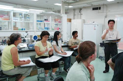
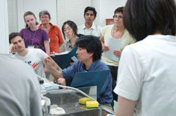
The first NIBB International Practical Course was held in 2007 with the aim of providing young scientists around the world with opportunities to learn cutting-edge experimental skills and to communicate with experts in the field. The course replaced our Bioscience Training Course, which had been held for twenty years and which had, for the most part, accepted participants from universities and institutes in Japan. The Third International Practical Course was held from June 30th to July 4th, 2008, focusing on techniques used in the biology of the moss Physcomitrella patens, which is an emerging model organism in plant biology.
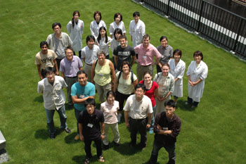
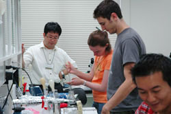
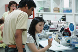
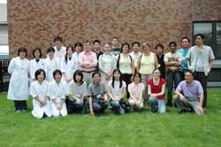
The eleven highly-qualified participants were from various countries: six from Japan, two from the U.S.A., and one each from Korea, Spain, and Sweden. They did excellent and enthusiastic work and enjoyed fruitful discussions throughout the five days of the course.
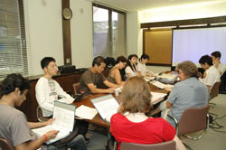
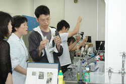
The course provided basic techniques including cultivation, observation of developmental processes at the cellular level, gene targeting, bioimaging, and bioinformatics. Drs. Andrew C. Cuming (Univ. Leeds), Yasuko Kamisugi (Univ. Leeds), Tetsuya Kurata (JST), Yoshikatsu Sato (JST), Yuji Hiwatashi (NIBB), Minoru Kubo (JST), Takashi Murata (NIBB), and Mitsuyasu Hasebe (NIBB) taught the course with the help of seven laboratory technicians. Several postdoctoral fellows introduced their research work using P. patens and shared detailed research procedures with the participants.
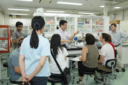
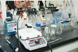
The course was given in a laboratory provided by NIBB and equipped with the necessary instruments and tools and with a good supply of material moss. We hope that the techniques and knowledge provided in this course will prove useful in the future work of all of the participants.
June 29, Sun
18:00 Get together
June 30, Mon
09:00 Welcome address
(Kiyotaka Okada, Director General of NIBB)
09:05 Introduction of course and Physcomitrella patens Hasebe and Cuming
09:35 Spore germination Hiwatashi
Culture and storage of protonemata and gametophores Hiwatashi
10:05 Observation of protonemata and gametophores Sato
Observation of protonema colony morphology Sato
11:05 Induction of gametangia and sporophytes Cuming and Kamisugi
Sporangium collection and storage Cuming and Kamisugi
Observation of antheridia and archegonia Cuming and Kamisugi
Observation of sperms Cuming and Kamisugi
12:05 Lunch
13:00 PEG-mediated transformation Hiwatashi, Cuming and Kamisugi
17:00 Crosses of mutants defective in the sporophyte formation Cuming and Kamisugi
18:00 Digital expression and microarray Kurata
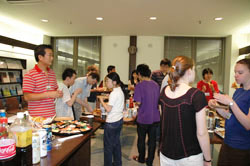
19:00 Welcome party
July 1, Tue
09:00 PEG-mediated transformation Hiwatashi, Cuming and Kamisugi
13:00 Lunch
14:00 Genomic DNA extraction Kubo
RNA extraction Kurata
Green PCR Kubo
15:00 DNA-gel-blot analysis Kubo
RNA interference Kubo
Gene disruption Kurata
16:00 Overexpression Kubo
17:00 Inducible expression Kubo
18:00 Dinner
19:00 Workshop 1: Targeting strategy Cuming and Kamisugi
July 2, Wed
09:00 PEG-mediated transformation Hiwatashi
10:00 GUS staining (1) Hiwatashi
11:00 PEG-mediated transformation Hiwatashi
12:00 Lunch
Free time
July 3, Thu
09:00 GUS staining (2) Hiwatashi
10:00 Observation of M-phase in a protonemal cell Hiwatashi
11:00 Observation of microtubules with indirect immunofluorescence microscopy Murata
Light and electron microscopy of protonemata embedded with epoxy resin Murata
12:00 Lunch
13:00 DAPI, Hoechst33342, and PI staining of protonemata Murata
Visualization of cell wall with calcoflur Murata
14:00 Live imaging Sato
15:00 Retrieving Genome Seqeuences for Gene targeting Cuming and Kamisugi
16:00 Insertion of reporter with gene targeting Sato
17:00 Cellular localization of fluorescent protein Sato
Protonema culture between agar-gelatin layers Sato
18:00 Dinner
19:00 Workshop 2: Bioimaging Sato, Murata, Cuming and Kamisugi
July 4, Fri
09:00 Measurement of DNA content with FACS Sato
11:00 Agrobacteria-mediated moss transformation:
Protonema and Agrobacteria Co-culture Kurata
12:00 Lunch
13:00 Agrobacteria-mediated moss transformation:
Protonema and Agrobacteria Co-culture Kurata
14:00 Hormone treatments Cuming and Kamisugi
15:00 Light response Sato
Gravitropic response Sato
16:00 Closing discussion All lecturers
17:00 Farewell party

