

Okazaki Large Spectrograph
About Okazaki Large Spectrograph(OLS)
Performance
・Light resource: 30kW Xenon arc lamp
・Spectroscopic method: Spectroscopy with a diffraction grating (90 cm×90cm)(250-1000 nm)
・Wavelength accuracy: ±2 nm(About 10 times that of general LED)
・Light intensity: Same level as sunlight in the south central area just below the equator. (light on the earth can be reproduced)
・Simultaneous multiwavelength experiments are possible.
Characteristic
・High wavelength accuracy.
From the above, it is possible to determine the action spectrum by precisely measuring the difference in effect depending on the wavelength of light. In addition, it can be applied to various researches as a powerful irradiation light source.
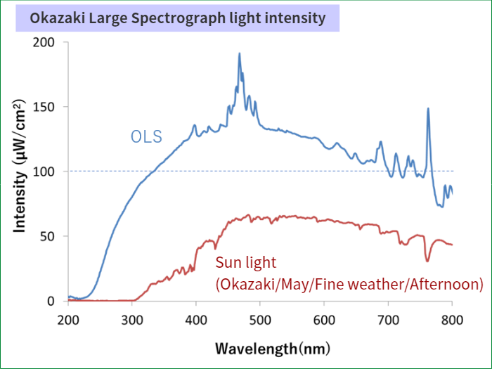
Fig.1. Okazaki Large Spectrograph light intensity.
Main composition
The spectroscopic condensing mirror (A7 in Fig. 2) is a 1m square, radius of curvature of 9 meters, F / 7 convex mirror, and the diffraction grating (A8 in Fig. 2) is about 15 cm square with 1200 grooves / mm. Blaze wavelength 250nm and 500nm double blazed plane. It is about 90 cm square with 36 diffraction gratings arranged in a mosaic pattern.
The distance from the center of the diffraction grating to the horizontal focal plane is about 10m.
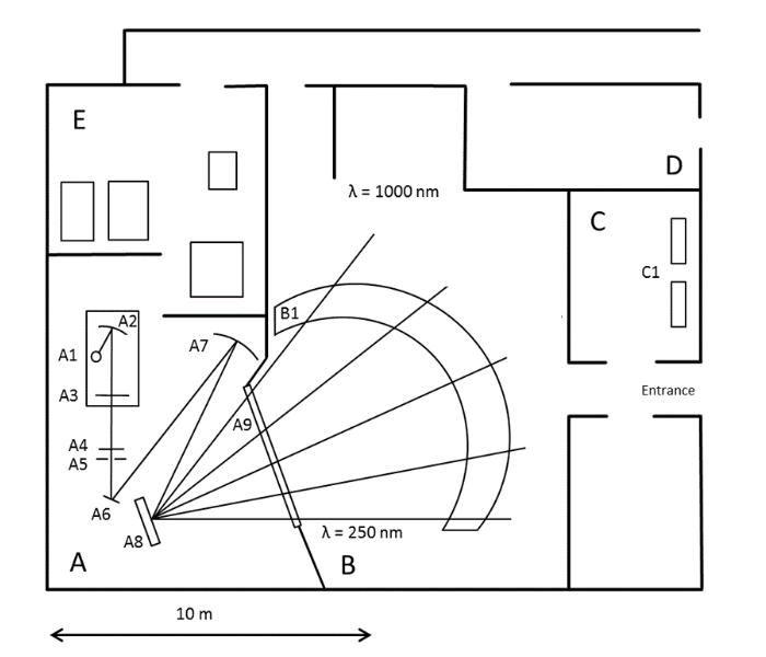
Fig.2. Okazaki Large Spectrograph configuration.
A: Light source/Spectral chamber, B: Irradiation room, C: Laboratory, D: Rest room, E: Power room, A1: 30kW xenon short arc lamp, A2: Light source condensing mirror, A3: Light source shutter, A4: Heat absorption shutter, A5: Light incident slit, A6: Deflection plane mirror, A7: Spectroscope condensing mirror, A8: Diffraction grating, A9: Stray light filter, B1: Focus curved table, C1: Incubator. (Modified the figure in Reference)
Irradiation method
There are methods such as direct irradiation, irradiation with a mirror and irradiation, and using a threshold box. Please contact us for more information.
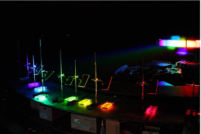
Fig.3. Illumination by shining light on a mirror.
The light intensity can be adjusted by changing the position and height of the mirror.
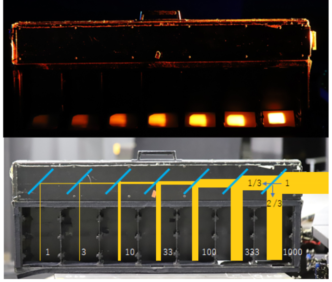
Fig.4. Threshold box.
The dimming mirror allows you to perform irradiation experiments with different light amounts at once. It is also possible to add a neutral density filter.
Facility introduction literature
1)Watanabe, M. (1995) Action spectroscopy: photomovement and photomorphogenesis spectra. CRC Handbook of Organic Photochemistry and Photobiology (W. M. Horspool and P-S Song eds.) CRC Press, Boca Raton, pp.1276-1288.
2)M. Watanabe, M. Furuya, Y. Miyoshi, Y. Inoue, I. Iwahashi and K. Matsumoto (1982) Design and performance of the Okazaki Large Spectrograph for photobiological research. Photochem. Photobiol. 36:491-498.
Other equipment related to spectral irradiation
A. Simple monochromatic light source
| A1. Hololight |
|---|
| LED compact monochromatic light irradiation device for 10 wavelengths. With a lens, it can diffuse and collect light. With strength adjustment device.(Available for rent. Please contact us for details. Simple instructions(Japanese only) |
| A2. LED irradiation array |
| Model Plant Research Facility, NIBB has an irradation device for 9 wavelengths. Advance reservation is possible. (Not available for rental, please contact us for details.) |
B. Photon meter
| B1. Photon meter(QTM-101) |
|---|
| The amount of light in the UV-visible near infrared region(200-1100nm)can be displayed as radiant energy value and photon density. It will be used in the OLS irradiation room. |
| B2. Solar spectroradiometer 【Hidamari-Mini】 |
| Located at Model Plant Research Facility, NIBB. The light source spectrum of 300-1100nm can be measured. |
| ※There is also a simple power meter (for microscopes). |
Recent major achievements
| Aihara Y, Maruyama S, Baird A H, Iguchi A, Takahashi S and Minagawa J. (2019) Green fluorescence from cnidarian hosts attracts symbiotic algae. PNAS 116, :2118-2123, press release (NIBB) |
|---|
| Kosugi M, Maruo F, Inoue T, Kurosawa N, Kawamata A, Koike H, Kamei Y, Kudoh S and Imura S. (2018) A comparative study of wavelength-dependent photoinactivation in photosystem II of drought-tolerant photosynthetic organisms in Antarctica and the potential risks of photoinhibition in the habitat. Ann Bot. 122, 1263-1278 |
| Matsuo M, Yoriko Ando Y, Kamei Y and Fukamachi S. (2018) A semi-automatic and quantitative method to evaluate behavioral photosensitivity in animals based on the optomotor response (OMR). Biology Open 7, bio033175 |
| Petroutsos D, Tokutsu R, Maruyama S, Flori S, Greiner A, Magneschi L, Cusant L, Kottke T, Mittag M, Hegemann P, Finazzi G and Minagawa J. (2016) A blue-light photoreceptor mediates the feedback regulation of photosynthesis. Nature, 537, 563-566, press release (NIBB) |
| Ikehata H, Higashi S, Nakamura S, Daigaku Y, Furusawa Y, Kamei Y, Watanabe M, Yamamoto K, Hieda K, Munakata N and Ono T. (2013) Action spectrum analysis of UVR genotoxicity for skin: The border wavelengths between UVA and UVB can bring serious mutation loads to skin. J. Invest. Dermatol. 133, 1850-1856. |
| Shikata T, Matsunaga S, Iseki M, Nishide H, Higashi S-I, Kamei Y, Yamaguchi M, Jenkinson I R and Watanabe M. (2013) Blue light regulates the rhythm of diurnal vertical migration in the raphidophyte red-tide alga Chattonella antiqua. J. Plankton Res. 35, 542-52. |
| Karahara I, Takaya E, Fujibayashi S, Inoue H, Weller J L, Reid J B and Sugai M. (2011) Development of the Casparian strip is delayed by blue light in pea stems. Planta, 234, 1019-1030. |
| Shikata T, Iseki M, Matsunaga S, Higashi S, Kamei Y and Watanabe M. (2011) Blue and red light-induced germination of resting spores in the red-tide diatom Leptocylindrus danicus. Photochem. Photobiol. 87, 590-597. |
| Arimoto-Kobayashi S, Sano K, Machida M, Kaji K and Yakushi K. (2010) UVA activation of N-dialkylnitrosamines releasing nitric oxide, producing strand breaks as well as oxidative damages in DNA, and inducing mutations in the Ames test. Mutat.Res. 691, 47-54. |