
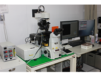
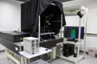
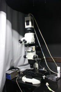
①Confocal and 2-photon microscope Leica TCS SP8 MP (Upright type)
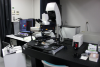
Place: Room No. 3-529, Yamate 3rd bldg. 5th Floor
Date of installation: Feb. 2013
Features:
Confocal imaging, Two-photon imaging (wavelength tunable), Z stack, Time-lapse, Photo stimulation, Spectral imaging (unmixing) and Photon counting
Specifications:
・Microscope: STP6000 (upright)
・Laser (nm): 488 / 552
・Two-photon laser: Wave length tunable 680-1300 nm (InSight DeepSee)
・Scanner: FOV scanner, Resonant scanner
・Internal detector for confocal and two-photon: MA PMT 1ch
・Non Descanned Detector (NDD) for two-photon imaging: HyD* 2ch
・Filter for NDD: DAPI/FITC, CFP/YFP, FITC/TRITC, FITC/TXR, TRITC/A633, mCherry/Cy5
 details(pdf link)
・Objective: x5, x25W (non-cover)
・Filter for visual observation: GFP-LP, DsRed-LP
 details (pdf link)
*HyD: High sensitivity Hybrid Detector

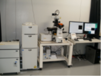
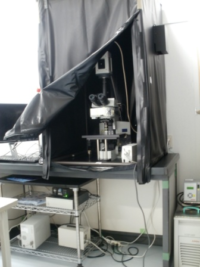
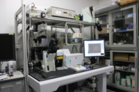
②Confocal and 2-photon microscope Leica TCS SP8 MP (Inverted type)
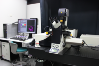
Place: Room No. 3-529, Yamate 3rd bldg. 5th Floor
Date of installation: Feb. 2013
Features:
Confocal imaging, Two-photon imaging (wavelength tunable), Z stack, Time-lapse, Photo stimulation, Large image (Tiling), Spectral imaging (unmixing) and Photon counting
Specification:
・Microscope:DMI6000CS (inverted)
・Laser (nm): 405 / 448 / 488 / 514 / 552
・Two-photon laser: tunable 680-1300 nm (InSight DeepSee)
・Scanner: FOV scanner, Resonant scanner
・Internal detector: MA PMT 1ch, HyD 3ch
・Non-Descanned Detector (NDD) for two-photon imaging: HyD 2ch
・Filter for NDD: DAPI/FITC, CFP/YFP, FITC/TRITC, FITC/TXR, TRITC/A633, mCherry/Cy5
 details (pdf link)
・Objective: x10, 20MI, x20W IR, x40W, (x40IR), x63W, (x63Oil)
・Filter for visual observation: GFP-LP, DsRed-LP
 details (pdf link)
・Stage Incubator1: CO2 chamber; RT~50℃, 5%CO2 (STXG-WSKMX-SET, TOKAI HIT)
・Stage Incubator2: Thermo Plate: 4-60℃ (TP-CHSQ-C, TOKAI HIT)
*HyD: High sensitivity Hybrid Detector

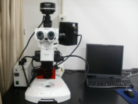
③Confocal and 2-photon (wavelength fixed) microscope Olympus FV1200 (Upright type)
Place: Room No. 3-532, Yamate 3rd bldg. 5th Floor
Date of installation: Dec. 2013
Features:
Confocal imaging, Two-photon imaging (wavelength fixed), Z stack, Time-lapse, Large image (Tiling)
Specifications:
・Microscope:BX61WIF (upright)
・Laser (nm): 473 / 559 / 635
・Two-photon laser: 1045 nm
・Scanner: Galvano scanner
・Detector for confocal imaging: MA PMT 3ch + GaAsP PMT 2ch
・Detector for 2-photon imaging: MA PMT 2ch+ GaAsP PMT 2ch
・Objective: x5, x10W (non-cover), x25W (non-cover)
The information of the objectives and the filters
 details
(pdf link)
details
(pdf link)

④Confocal and 2-photon (wavelength fixed) microscope Olympus FV1200 (Inverted type)
Place: Room No. 3-532, Yamate 3rd bldg. 5th Floor
Date of installation: Dec. 2013
Features:
Confocal imaging, Two-photon imaging (wavelength fixed), Z stack, Time-lapse, Large image (Tiling)
Specifications:
・Microscope: IX83 (inverted)
・Laser (nm): 405 / 473 / 559 / 635
・Two-photon laser: 1045 nm
・Scanner: Galvano scanner
・Detector for confocal imaging: MA PMT 3ch + GaAsP PMT 2ch
・Detector for 2-photon imaging: GaAsP PMT 2ch
・Objective: x10, x20dry, x40dry, x60S
The information of the objectives and the filters
 details
(pdf link)
details
(pdf link)

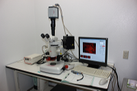
⑤ Confocal microscope Nikon A1R
Place: Room No. B36, NIBB
Date of installation: Mar. 2010
Features: Confocal imaging, Z stack, Time-lapse, Photo stimulation and Large image (Tiling)
Specifications:
・Microscope: Ti-E (inverted)
・Laser (nm): 405 / 457 / 488 / 514 / 561 / 633
 Details of Lasers, 1st dichroic mirrors and emission filters (pdf link)
・Scanner: Galvano scanner, Resonant scanner
・Detector: MA PMT 2ch + GaAsP PMT 2ch (Ch2 and Ch3: GaAsP)
・Objective: (x4), x10, 20, (x20MI), x40W, (x40dry), x60W, x60Oil, x100oil
 details (pdf link)
・Fluorescent filter for visual observation: DAPI, FITC-LP, TxRed, CFP, (FITC-BP)
 details(pdf link)
・Stage incubator: RT~50℃, 5%CO2 (INU-TIZ-F1, TOKAI HIT)
・Piezo stage: (range: ~60μm)

⑥Confocal microscope Nikon A1Rsi
Place: Room No. 3-531, Yamate 3rd bldg. 5th Floor
Date of installation: Mar. 2010
Features:
Confocal imaging, Z stack, Time-lapse, Photo stimulation, Spectral imaging (unmixing) and Large image (Tiling)
Specifications:
・Microscope: Ti-E (inverted)
・Laser (nm): 405 / M-Ar; 457, 488, 514 / 561 / 633
 details(
pdf link)
details(
pdf link)
・Scanner: Galvano scanner, Resonant scanner
・Internal Detector: MA PMT 2ch + GaAsP PMT 2ch
・Non-Descanned Detector (NDD): HyD 2ch
・Objective: x10, x20, x40W, (x40dry), x60W, x60Oil
 details (pdf link)
・Fluorescent filter for visual observation: DAPI, FITC-LP, TxRed, CFP
 details (pdf link)

⑦Confocal microscope Olympus FV1000
Place: Room No. 3-529, Yamate 3rd bldg. 5th Floor
Date of installation: Mar 2006
Features:
Confocal imaging, Z stack, Time-lapse, Photo stimulation, Spectral imaging (unmixing) and Large image (tiling)
Specifications:
・Microscope: BX61 (upright)
・Laser (nm): 405 / M-Ar; 458, 488, 515 / 543 / 633
・Scanner: Galvano scanner
・Detector: MA PMT 4ch
・Objective: x10, x20, x20W (non-cover), x40W, x40Oil, x60Oil
 The details of the objectives and the filters (pdf link)

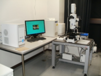
⑧ CSU-X1 Spinning Disc Confocal Microscope
Place: Room No. B65, NIBB
Date of installation: Mar. 2012
Features:
Spinning disc confocal, Z stack, Time-lapse, multi point imaging
Specifications:
・Microscope: Olympus IX71 (Inverted and manual type)
・Spinning disc: Yokogawa CSU-X1
・Camera: ImagEM 1K, OrcaFlash 4.0
・Laser (nm) : 405 / 488/ 561 / 640
・Objective: x10, x20, x30S, x60W, x60S
・XY motor stage:
・Z piezo stage: range 350μm
・Z capra
・Software: Metamorph
・Stage Incubator: RT~50℃, 5%CO2 (INUBSF-FI-RK, TOKAI HIT)

⑨Olympus 2-photon microscope FVMPE-RS
Place: Room No. B65, NIBB
Date of installation: Dec. 2013
Features:
Two-photon imaging (2 laser line: wavelength tunable and 1040nm fixed), Z-stack, Time-lapse
Specifications:
・Microscope: BX63LF (Upright)
・Laser: tunable 680-1300 nm (InSight DeepSee), 1040nm line
・Objective: x10, x25W IR (non-cover), x40W
 details (pdf link)

⑩Automated Fluorescence Microscope Olympus BX63
Place: Room No. B36, NIBB
Date of installation: Feb. 2011
Features:
Fluorescence imaging, DIC, Z-stack, Time-lapse, Large image (Tiling)
Specifications:
・Microscope: BX63 (upright)
・Software: cellSens
・Objective: x10, x20, x40, x60Oil, x100Oil
・Filter: DAPI, GFP-BP, GFP-LP, DsRed
 details (pdf link)

⑪ Fluorescent Microscope Keyence BZ8000 (inverted)
Place: Room No. B10, NIBB common bldg.
Date of installation: Feb. 2007
Features: Multi color imaging
Specification:
・Objective: x20, x60Oil
・Filter: DAPI-B, GFP-B, GFP-L, TxRed
 details (pdf link)

⑫ Luminescence Microscope Olympus BX61WI
Place: Room No. B36, NIBB
Date of installation: Feb. 2007
Features: Luminescence and weak fluorescence imaging, Time-lapse
Specifications:
・Microscope: BX61WI (upright)
・Camera: ImagEM C9100-13 (512pix), shared with MVX10
・Objectives: x10, x20W (non-cover)
・Filiters: GFP-B, RFP-L
・Software: Metamorph
 details (pdf link)

⑬ Macro zoom microscope Olympus MVX10
Place: Room No. B36, NIBB
Date of installation: Sep. 2013
Features: Luminescence and weak fluorescence imaging, Time-lapse
Specifications:
・Microscope: Macrozoom MVX10
・Camera: ImagEM C9100-13 (512pix), shared with BX61WI
・Objective: MVPLAPO 1x (N.A. 0.25), MVPLAPO 2x (N. A. 0.5)
・Filiters: GFP-BP, RFP-BP
・Software: Metamorph

⑭ Fluorescent stereo microscop Leica M205FA
Place: Room B66, NIBB
Date of installation: Mar. 2011
Features:
・Multi color imaging, Z-stack, Time-lapse
Specifications:
・Camera: DFC360FX (monochrome), DFC550(color)
・Objectives: x1, x5
・Filter: DAPI, GFP2 (LP), GFP3 (BP), DSR, YFP

⑮ Fluorescent stereo microscope Olympus SZX16(Myodaiji)
Place: Room No. B66, NIBB
Date of installation: Dec. 2010
Specifications:
・Objectives: x0.5, x1, x2
0.5X: SDFPLAPO 0.5XPF (N.A. 0.075, W.D. 70.5 mm)
1X: SDFPLAPO 1XPF (N.A. 0.15, W.D. 60 mm)
2X: SDFPLAPO 2XPFC(N.A. 0.3, W.D. 20 mm)
・Zoom ratio: 16.4 (0.7x~11.5x)
・Fluorescent filter: GFP-HQ, GFP, RFP1
GFP-HQ Ex: BP460-480, Em: BA495-540
GFP Ex: BP460-495, Em: BA510IF
RFP1 Ex: BP530-550, Em: BA575IF
・Camera: DP71
・Software: DP controller, DP manager

⑯ Fluorescent stereo microscope Olympus SZX16 (Yamate)
Place: Room No. 3-529, Yamate 3rd bldg. 5th Floor
Specifications:
・Fluorescent filter:
GFP Ex: BP460-495, Em: BA510IF
GFP-HQ Ex: BP460-480, Em: BA495-540
RFP1 Ex: BP530-550, Em: BA575IF
・Software:cell Sens

⑰ Fluorescent stereo microscope Leica MZFLII
Place: Room No. B36, NIBB
Specifications:
・Fluorescent filter:
GFP2 (LP): Ex: 480/40, DM: 505, Em: 510LP
GFP3 (BP): Ex: 470/40, 495, Em: 525/50
DsRed: Ex: 546/10, DM: 565, Em: OG590
・Bright Field: Transmitted illumination, Epi-illumination
・Camera:3CCD degital, modelC7780-20

⑱ Tabletop microscope Hitachi Miniscope TM4000Plus
Place: Room No. B36, NIBB
Date of installation: Dec. 2017
Feature: Cool stage

⑲ Macro confocal microscope Nikon AZ-C1
Place: Room No. 3-531, Yamate 3rd bldg. 5th Floor
Date of installation: Mar. 2010
Specifications:
・Laser (nm): 488, 561
・Objectives: x1, x2, x5
・Zoom: x1~x8

⑳ IR-LEGO
Place: Room B66, NIBB
Date of installation: Mar. 2010
Feature: IR-LEGO (Infrared laser-evoked gene operator system) is the system to induce gene expression of local area in living cells using IR laser irradiation.
(Ref:Kamei et al. Nat. Methods 6, 79-81. 2009,(Pubmed)Deguchi et al. Dev. Growth Differ. 51, 769-775. 2009(Pubmed))。
Specification:
・Microscope: IX81 (inverted)
・IR laser: 1440-1480nm, 1W (HPU 10X09-001, Furukawa)
・Objectives: x20 and x40 (for IR irradiation), x4, x10, x20 x40 for Observation
・Filter: DAPI, GFP-BP, GFP-LP, RFP
・Camera: DP72
・Software: cellSens

㉑ Rigaku R_mCT2
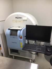
Place: Room 32-33, NIBB
Date of installation: Feb. 2013
Specification:
・High-speed shooting: Up to 17 seconds
・Low exposure (10mGy or less in the fastest 17 seconds)
・High resolution:10mm (FOV 5mm)
・Respiratory / heartbeat synchronization function

㉒ WorkStation 1 / Myodaiji
Place: Room B36, NIBB
Date of installation: Feb. 2018
Specifications:
・OS: Windows 10 64bit
・Intel® Xeon E5-2687W v4 @3.00GHz 3.00GHz (2 processor)
・RAM: 64GB
・GPU: 12GB (NVIDIA TITAN X)
・Software: NIS-elements, LAS-X (including FLIM and FCS option), FIJI, Office

㉓ WorkStation 2 / Myodaiji
Place: Room B36, NIBB
Date of installation: Feb. 2019
Specifications:
・OS: Windows 10 64bit
・Intel® Xeon 8-core W-2145 3.7GHz
・RAM: 128GB
・GPU: 16GB (NVIDIA Quadro P5000)
・Software: Imaris suite (Including Filament tracer and CELL option), Huygens, Metamorph*, FIJI, Office
*Metamoph needs the license key.

㉔ WorkStation 3 / Yamate
Place: Room No. 3-531, Yamate 3rd bldg. 5th Floor
Date of installation: Feb. 2019
Specifications:
・OS: Windows 10 64bit
・Intel® Xeon 8-core W-2145 3.7GHz
・RAM: 128GB
・GPU: 16GB (NVIDIA Quadro P5000)
・Software: Imaris suite, NIS-elements, LAS-X, FIJI , Office

㉕ WorkStation 4 / Yamate
Place: Room No. 3-531, Yamate 3rd bldg. 5th Floor
Date of installation: Feb. 2013
Specifications:
・OS: Windows 7 64bit
・Intel® Xeon® CPU E5-2650 0 @ 2.00GHz 2.00GHz
・RAM; 16GB
・GPU: 1GB (NVIDIA GeForce480)
・Software: Hyvolution, LAS-AF (no 3D function), FIJI (ImageJ)

㉖ WorkStation 5 / Myodaiji
Place: Room B32-33, NIBB
Date of installation: Dec. 2019
Specifications:
・Model: iMacPro
・OS: MacOS 10.14.6
・3.2GHz 8-Core Intel Xeon W processor
・RAM: 32GB 2,666MHz DDR4
・GPU: Radeon Pro Vega 64
・Software: OsiriX, Fiji (ImageJ)

㉗ WorkStation 6 / Myodaiji
Place: Room B32-33, NIBB
Date of installation: Dec. 2013
Specifications:
・Model: MacPro (Mid 2012)
・OS: X Yosemite ver. 10.10.5
・2 x 2.4GHz 6-Core Intel Xeon processor
・RAM: 16 GB 1,333MHz DDR3 ECC
・GPU: ATI Radeon HD 5870 1024MB
・Software: OsiriX, Fiji (ImageJ)