
タヌキノショクダイ Thismia abei: Cropped from a photo by Kenji Suetsugu, Suetsugu et al. (2026), CC BY 4.0; ヒナノボンボリ Oxygyne hyodoi: Cropped from a photo by Syozi Hyodo, Cheek et al. (2018) Fig. 6, CC BY 4.0
タヌキノショクダイ科
Thismiaceae
タヌキノショクダイ科 Thismiaceaeは、ハプロティスミア属 Haplothismia (1種)、ヒナノボンボリ属 Oxygyne (6種)、ムジナノショクダイ属 Relictithismia (1)、タヌキノショクダイ属 Thismia (117種)、チプチニア属 Tiputinia (1種)の5属が知られている(POWO)。
The family Thismiaceae comprises five genera: Haplothismia (one species), Oxygyne (six species), Relictithismia (one species), Thismia (117 species), and Tiputinia (one species) (POWO).

Distribution of the genera of Thismiaceae
Source: POWO (2026). Plants of the World Online. Facilitated by the Royal Botanic Gardens, Kew. Published on the Internet; https://powo.science.kew.org/. Retrieved 3 June 2026. Distribution data from Kew Backbone Distributions (WCVP). © World Checklist of Vascular Plants, licensed under CC BY 3.0.
タヌキノショクダイ科各属の分布
Distribution of the genera of Thismiaceae
タヌキノショクダイ科は、全種が菌従属栄養性で光合成を行わず、熱帯を中心に温帯までの暗い林床に生育する。POWOの分布図は国単位で分布を示しているため、実際の分布域よりも広く表示されている。
All species of Thismiaceae are mycoheterotrophic and non-photosynthetic, occurring on the dark forest floor from tropical to temperate regions, with their center of diversity in the tropics. The distribution maps in POWO are based on country-level records and therefore depict broader distributions than the actual species ranges.
Natural habitat of Thismia kobensis
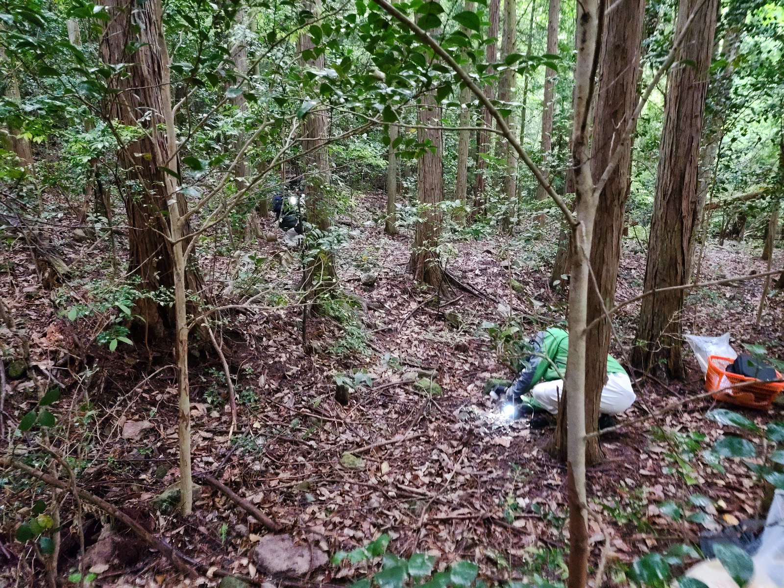
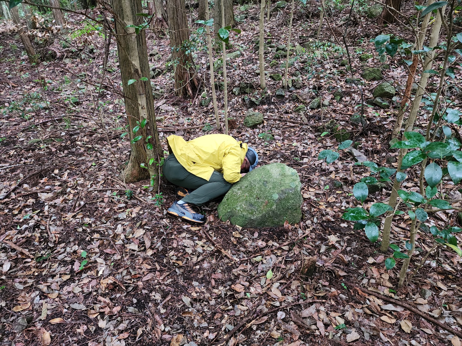
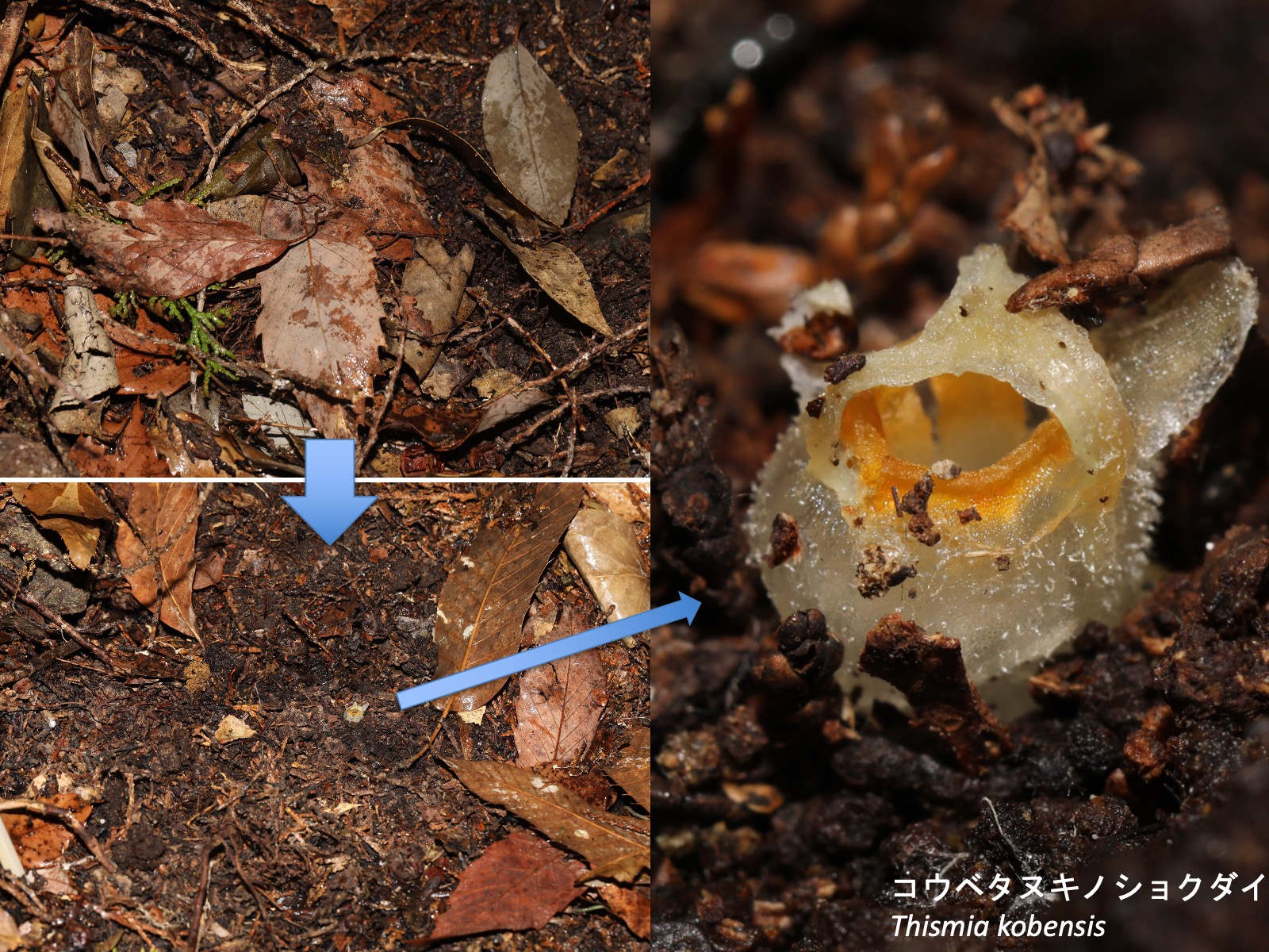
ヒノキの植林にコナラなどの広葉樹が侵入した林内で、コウベタヌキノショクダイの生育地点周辺には他の草本植物はほとんど生育していない。6月に写真の人物がいる場所で昨秋の落葉を取り除くと、その下で開花している個体が観察された。The photographs above show a natural habitat of Thismia kobensis. The site is located in a Japanese cypress (Chamaecyparis obtusa) plantation that has been slightly invaded by broad-leaved trees such as Quercus serrata. Few other herbaceous plants occur in the vicinity of the T. kobensis population. When the leaf litter deposited during the previous autumn was removed at the location indicated by the person in the photographs in June, flowering individuals were found beneath it.
タヌキノショクダイ科に特有の形質
Distinctive characters of Thismiaceae
タヌキノショクダイ科は、 (1) 全種が完全菌従属栄養性であること、(2) ガク片、花弁、雄ずいの少なくとも1つに異所的な突起が形成されること (Haplothismiaを除く)、という特徴がある。
Thismiaceae is characterized by two shared characters: (1) all species are fully mycoheterotrophic, and (2) ectopic appendages are formed on at least one of the sepals, petals, or stamens (except in Haplothismia).
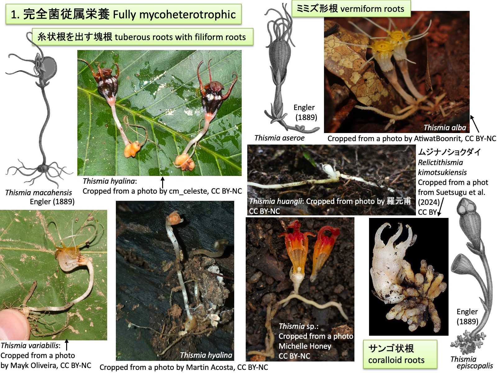
Fully mycoheterotrophic
Thismia macahensis, Thismia aseroe, Thismia (= Bognisia) episcopalis: Drawings are reproduced from Engler (1889); Thismia variabilis: Cropped from a photo by Mayk Oliveira, https://www.inaturalist.org/photos/595106823, CC BY-NC; Thismia hyalina: Cropped from a photo by cm_celeste, https://www.inaturalist.org/photos/468954877, CC BY-NC; Thismia hyalina:_Cropped from a photo by Martin Acosta, https://www.inaturalist.org/photos/126745926, CC BY-NC; Thismia huangii: Cropped from a photo by 羅元甫, https://www.inaturalist.org/photos/283029670, CC BY-NC; Thismia alba: Cropped from a photo by AtiwatBoonrit, https://www.inaturalist.org/photos/138703569, CC BY-NC; Thismia sp.: Cropped from a photo by Michelle Honey, https://www.inaturalist.org/photos/65865470, CC BY-NC; Relictithismia kimotsukiensis: Cropped from a photo from Suetsugu et al. (2024), CC BY 4.0
1. 完全菌従属栄養
Fully mycoheterotrophic
タヌキノショクダイ科Thismiaceaeの全種は完全菌従属栄養性 fully mycoheterotrophicである(塚谷 2016; 末次 2023)。多くの植物は複数種のアーバスキュラー菌根菌と共生し、光合成によって生産した炭水化物や脂質を菌類に供給する代わりに、菌類からリン、窒素、その他無機塩や水分を得ている(Parniske 2008)。一方、タヌキノショクダイ科の植物は光合成を行わないため、菌類に光合成産物を供給せず、生活に必要な炭素源を含む栄養分と水分を菌類から得ている(Gomes et al., 2017; Merckx et al., 2017)。菌類が利用する炭素源はもともと周辺の光合成植物に由来するため、タヌキノショクダイ科の植物は菌類を介して周辺の植物から間接的に炭素を獲得しているともいえる。共生するアーバスキュラー菌根菌は通常1種または少数種に限られ(Guo et al. 2019、Gomes et al. 2017)、周辺の光合成植物と比較して宿主特異性が高く、共生関係の特殊化が進んでいると考えられている(Gomes et al. 2017)。
タヌキノショクダイ科では、糸状根を出す塊根 tuberous roots with filiform roots、ミミズ形根 vermiform roots、サンゴ状根 coralloid rootsなどの根形態が知られている(Feller et al. 2022)。このような根形態の多様化は、(1) 菌糸が共生できる組織を増やすこと、(2) 広い表面積によって菌糸との接触、感染機会を高めること、(3) 菌由来の栄養分を短距離で効率よく植物体へ輸送すること、という三つの条件を最適化する方向に進化した結果であり、各タイプの根がそれぞれ異なる最適解に到達したものと考えられている(Feller et al. 2022) 。上図では、T. macahensis、T. hyalina、T. variabilisが塊根型、T. aseroe、T. alba、T. huangii,T. sp.がミミズ形根型、T. episcopalisおよびRelictithismia kimotsukiensisはサンゴ状根型である。
All species of Thismiaceae are fully mycoheterotrophic. Most plants form symbiotic associations with multiple species of arbuscular mycorrhizal fungi, supplying the fungi with carbohydrates and lipids produced by photosynthesis in exchange for phosphorus, nitrogen, other mineral nutrients, and water (Parniske 2008). In contrast, because members of Thismiaceae are non-photosynthetic, they do not provide photosynthates to their fungal partners but instead obtain their nutrients and water, including carbon sources required for growth and survival, from the fungi (Gomes et al. 2017; Merckx et al. 2017). Since these carbon sources ultimately originate from surrounding photosynthetic plants, members of Thismiaceae can be regarded as indirectly acquiring carbon from neighboring plants via their fungal associates. The arbuscular mycorrhizal fungi associated with Thismiaceae are usually restricted to one or a few species (Guo et al. 2019; Gomes et al. 2017), exhibiting greater host specificity than those associated with surrounding photosynthetic plants, and are therefore considered to represent highly specialized symbiotic relationships (Gomes et al. 2017).
Three major root morphologies are recognized in theThismiaceae: tuberous roots with filiform roots, vermiform roots, and coralloid roots (Feller et al. 2022). The diversification of these root types has been hypothesized to reflect optimization of three functional requirements: (1) increasing the amount of tissue available for fungal colonization, (2) enhancing opportunities for fungal contact and infection through a larger surface area, and (3) facilitating efficient transport of fungus-derived nutrients to the plant body over short distances. The different root types are therefore thought to represent alternative evolutionary solutions to these functional demands (Feller et al. 2022). In the figure above, T. macahensis, T. hyalina, and T. variabilis possess tuberous roots; T. aseroe, T. alba, T. huangii, and Thismia sp. possess vermiform roots; and T. episcopalis and Relictithismia kimotsukiensis possess coralloid roots.
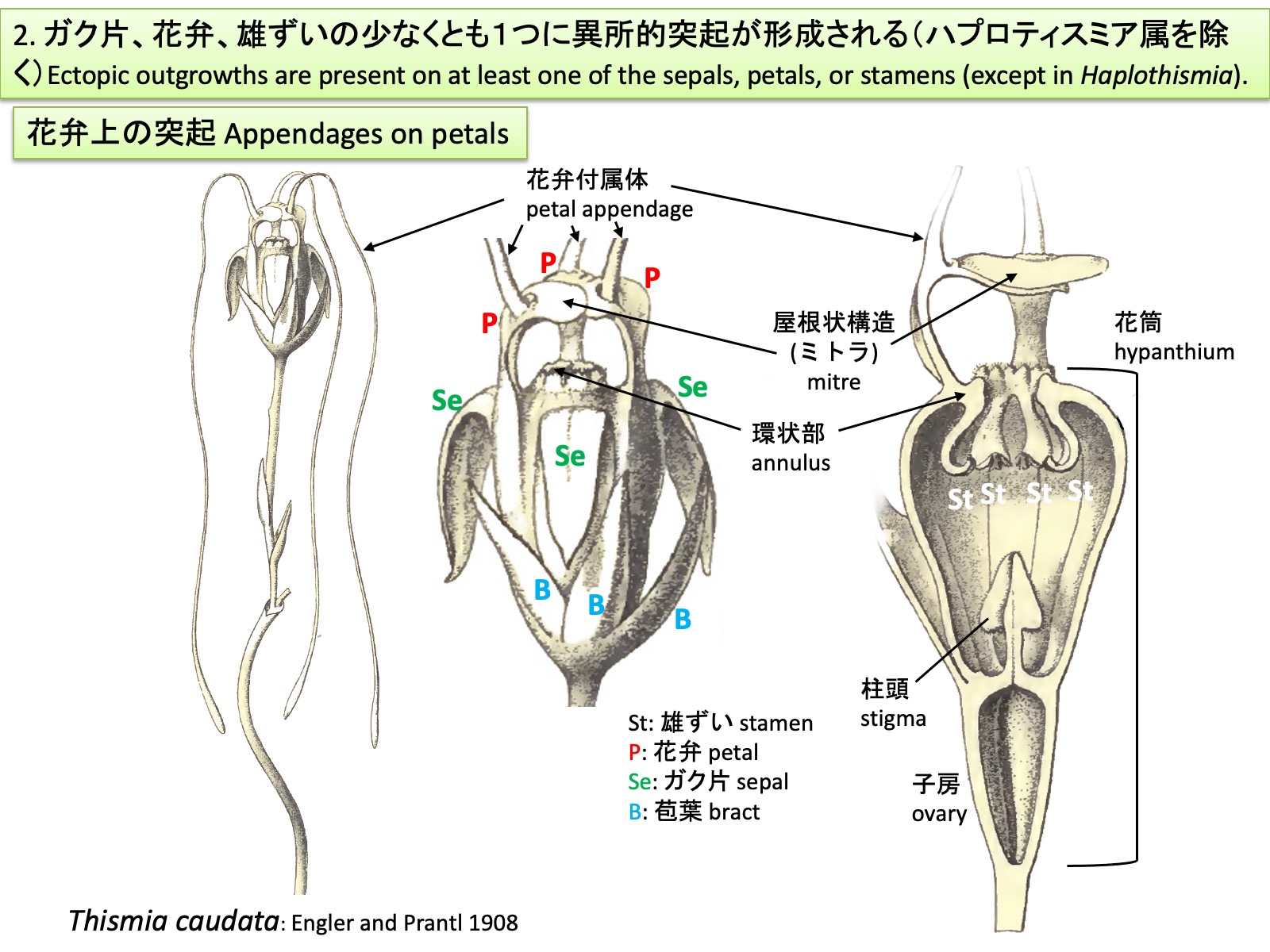
2. Ectopic outgrowths are present on at least one of the sepals, petals, or stamens (except in Haplothismia)
Thismia caudata: Drawings are reproduced from Engler and Prantl (1908)
2. ガク片、花弁、雄ずいの少なくとも1つに異所的突起が形成される(ハプロティスミア属を除く)
Ectopic outgrowths are present on at least one of the sepals, petals, or stamens (except in Haplothismia).
タヌキノショクダイ科の花も他の被子植物の花と同様に、外側からガク片、花弁、雄ずい、雌ずいが形成される。ガク片は外花被、花弁は内花被とも呼ばれる。上図のブラジル産Thismia caudataでは、花弁から糸状の花弁付属体petal appendages が伸長するとともに、3枚の花弁の先端側が合着して屋根状構造 mitreを形成する(Engler and Prantl 1908; Ríos and da Cruz 2023)。Mitreはラテン語mitraに由来する英語表記で、日本語ではミトラと呼ばれることが多い。ガク片、花弁および雄ずいは癒合して花筒を形成し、その開口部は隆起して環状部annulusとなる。雄ずいは花筒内に下垂し、葯は花筒の外側に向かって裂開する。
Flowers of Thismiaceae, like those of other angiosperms, are composed of sepals, petals, stamens, and carpels arranged from the outside inward. The sepals are also referred to as the outer perianth, and the petals as the inner perianth. In the Brazilian species Thismia caudata, shown above, filamentous petal appendages elongate from the petals, while the distal portions of the three petals fuse to form a roof-like structure known as a mitre (Engler and Prantl 1908; Ríos and da Cruz 2023). The term “mitre” is derived from the Latin mitra and is often rendered in Japanese as mitora. The sepals, petals, and stamens are fused to form a floral tube, the opening of which is elevated into an annulus. The stamens hang down within the floral tube, and the anthers dehisce outward, away from the center of the floral tube.

Appendages on sepals
Thismia megalongensis: Cropped from a photo by izakschoon, https://www.inaturalist.org/photos/176122303, CC BY-NC
ガク片上の突起
Appendages on sepals
Thismia megalongensisでは、T. caudataと同様に、花弁付属体(水色矢印)と屋根状構造(黄緑色矢印)が形成される。さらに、ガク片からも突起(ガク片付属体:黄色矢印)が形成される。 In Thismia megalongensis, as in T. caudata, petal appendages (light blue arrows) and a mitre (light green arrows) are present. In addition, the sepals bear appendages (sepal appendages; yellow arrows).

Appendages on stamens_1
Thismia appendiculata: Drawings are reproduced from Schlechter (1919)
雄ずい上の突起_1
Appendages on stamens_1
タヌキノショクダイ属の多くの種では、雄ずいの葯隔や花糸が伸長し、雄ずい同士が癒合したり、先端側が膜状に広がったり、伸長した先端に腺を形成したりする。上図のThismia appendiculataでは、6本の雄ずいが癒合して筒状になる。右上図では、1本の雄ずいを水色で示した。右下図では葯を黄色で示した。
In many species of Thismia, the connective tissue and filaments of the stamens become elongated, resulting in fusion between adjacent stamens, expansion of their distal portions into laminar structures, or the formation of glands at the tips of the elongate structures. In Thismia appendiculata, shown above, the six stamens are fused to form a tube. In the upper right figure, a single stamen is highlighted in light blue. In the lower right figure, the anthers are shown in yellow.
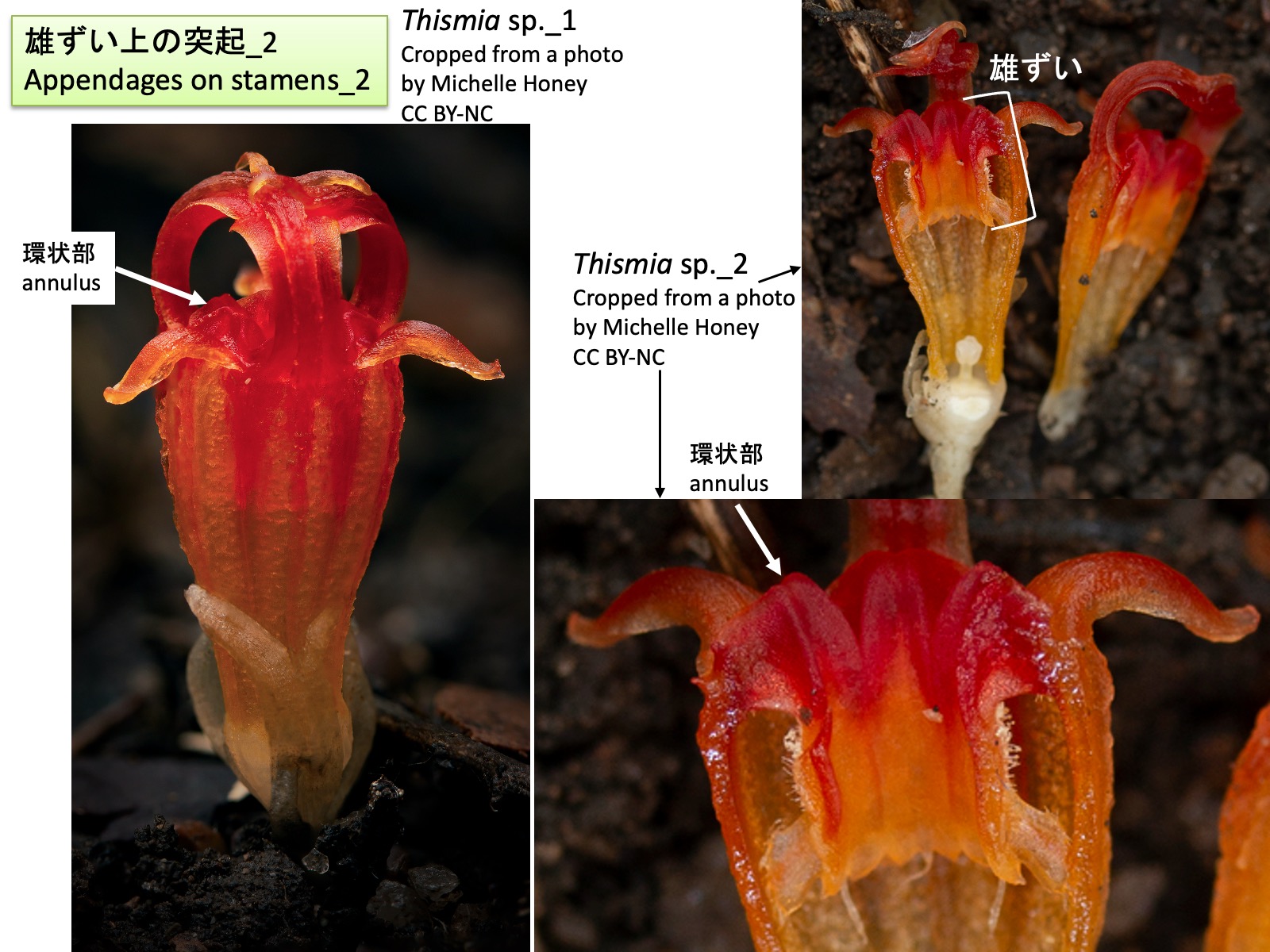
Appendages on stamens_2
Thismia sp._1: Cropped from a photo by Michelle Honey, https://www.inaturalist.org/photos/65865480, CC BY-NC; Thismia sp._2: Cropped from a photo by Michelle Honey, https://www.inaturalist.org/photos/65865470, CC BY-NC
雄ずい上の突起_2
Appendages on stamens_2
タヌキノショクダイ属の種では、上図のThismia sp.のように、雄ずいの花糸基部が伸長して環状部 annulus を形成する。ムジナノショクダイ属Relictithismiaにおいても、環状部が認められるが(Suetsugu et al. 2024)、それがタヌキノショクダイ属と同様に雄ずいの花糸基部に由来するかどうかについては、発生過程がまだ報告されていない。
In species of Thismia, as in Thismia sp. shown above, the basal portions of the stamen filaments elongate to form the annulus. An annulus is also present in Relictithismia (Suetsugu et al. 2024), but its developmental process has not yet been reported, and it remains unclear whether it is derived from the basal portions of the stamen filaments, as in Thismia.
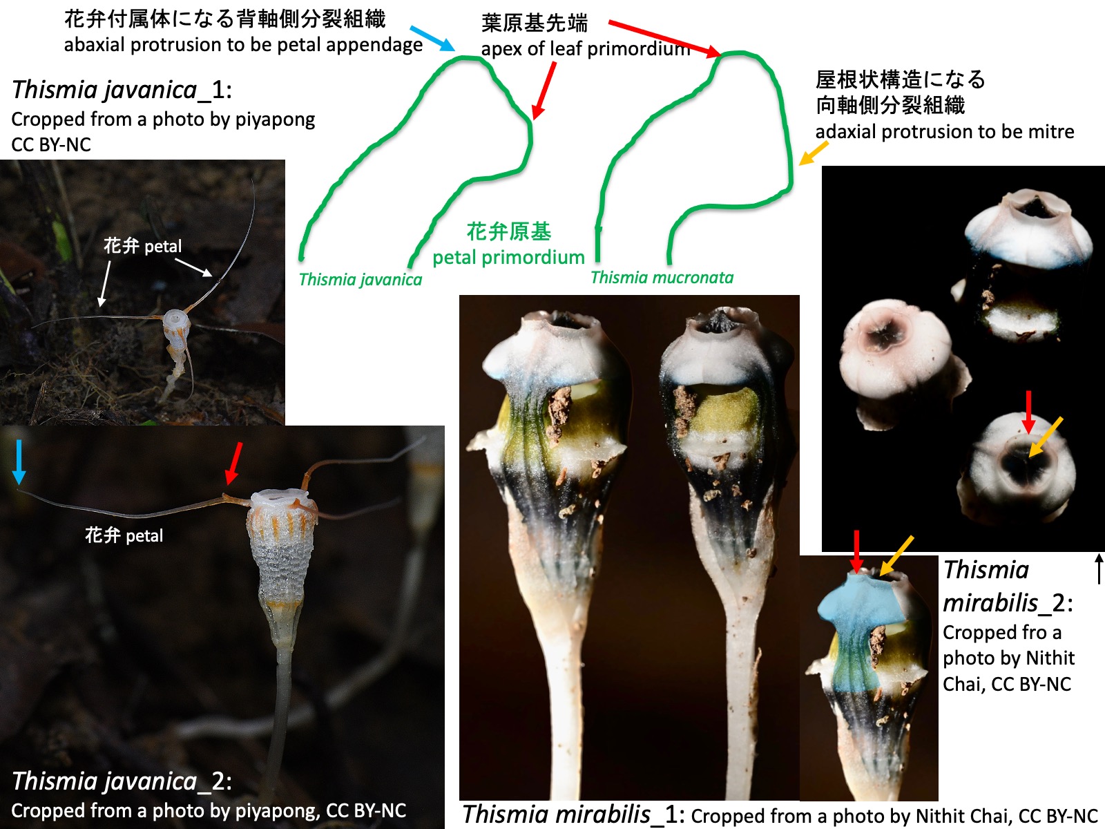
Development of petal appendages, the mitre, and the annulus
Thismia javanica_1: Cropped from a photo by piyapong, https://www.inaturalist.org/photos/441606101, CC BY-NC; Thismia javanica_2: Cropped from a photo by piyapong, https://www.inaturalist.org/photos/441609408, CC BY-NC; Thismia mirabilis_1: Cropped from a photo by Nithit Chai, https://www.inaturalist.org/photos/304715542, CC BY-NC; Thismia mirabilis_2: Cropped from a photo by Nithit Chai, https://www.inaturalist.org/photos/304715575, CC BY-NC
花弁付属体、屋根状構造、環状部の発生
Development of petal appendages, the mitre, and the annulus
ガク片、花弁、雄ずいの突起はどのように形成されるのだろうか。Nuraliev et al. (2021)は東南アジア産のThismia annamensis、T. javanica、T. mucronataの花発生過程を観察し、以下のことを報告した。
(1) T. javanicaでは、花弁原基の発生過程において、花弁原基の背軸側に新たな分裂組織が形成され、それが伸長して花弁付属体となる。上図左側にT. javanicaの花弁原基の模式図を示す。T. javanica_2の写真の青色矢印は花弁付属体の先端部を示している。一方、赤色矢印で示した突起についてはNuraliev et al. (2021)では言及されていない。この突起は花弁原基の先端部が伸長したものかもしれないが、その起源を明らかにするためには発生過程の観察が必要である。
(2) T. mucronataでは、花弁原基の発生過程において、花弁原基の向軸側に新たな分裂組織が形成され、それが伸長、癒合して屋根状構造となる。上図右側にT. mucronataの花弁原基の模式図を示す。Nuraliev et al. (2021)はT. mucronataを観察したが、上図にはShepeleva et al. (2020)においてT. mucronataの姉妹種とされ、類似した花形態をもつT. mirabilis の花の写真を示した。 右下の写真で青色に着色した部分は1枚の花弁に相当する。花弁先端部には隆起が認められ(赤色矢印)、さらにその先へ伸長している(黄色矢印)。赤色矢印で示した部分が花弁原基の先端部、黄色矢印で示した部分が向軸側に形成された分裂組織に由来する組織であるようにも見える。しかし、Nuraliev et al. (2021)では屋根状構造の形成過程そのものは観察されておらず、この点についてもさらなる発生学的研究が必要である。
(3) 雄ずい原基は花筒基部側に向かって伸長していた。雄ずいの花糸基部の背軸側は伸長して環状部を形成していた。また、葯隔や花糸が異所的に伸長することによって複雑な形態の雄ずいが形成されていた。
これらの観察結果から、タヌキノショクダイ科に見られる特異な花形態の少なくとも一部は、花弁原基、ガク片原基、花糸および葯隔に異所的な分裂組織が形成され、それらが伸長することによって生じたものと考えられる。
How are the appendages of the sepals, petals, and stamens formed? Nuraliev et al. (2021) investigated floral development in the Southeast Asian species Thismia annamensis, T. javanica, and T. mucronata and reported the following observations.
(1) In T. javanica, during petal development, a new meristematic region is formed on the abaxial side of the petal primordium and subsequently elongates to form a petal appendage. A schematic diagram of the petal primordium of T. javanica is shown on the left side of the figure above. The blue arrow in photograph T. javanica_2 indicates the tip of a petal appendage. In contrast, the projection indicated by the red arrow was not mentioned by Nuraliev et al. (2021). This structure may represent an elongation of the distal portion of the petal primordium, but observations of its developmental process are required to clarify its origin.
(2) In T. mucronata, during petal development, a new meristematic region is formed on the adaxial side of the petal primordium. This tissue elongates and fuses to form a mitre. A schematic diagram of the petal primordium of T. mucronata is shown on the right side of the figure above. Although Nuraliev et al. (2021) studied T. mucronata, the figure presented here shows flowers of T. mirabilis, which was recovered as the sister species of T. mucronata by Shepeleva et al. (2020) and possesses a similar floral morphology. In the lower right photograph, the blue-colored region corresponds to a single petal. A prominent swelling is present near the petal apex (red arrow), and an elongate structure extends beyond it (yellow arrow). The region indicated by the red arrow appears to correspond to the distal portion of the petal primordium, whereas the structure indicated by the yellow arrow may be derived from the meristematic tissue formed on the adaxial side. However, the developmental stages leading to mitre formation were not observed by Nuraliev et al. (2021), and further developmental studies are needed to test this interpretation.
(3) Stamen primordia elongated toward the base of the hypanthium. The abaxial side of the basal portion of the staminal filaments elongated to form the annulus. In addition, ectopic elongation of the connective tissue and filaments gave rise to the complex morphology of the stamens. These observations suggest that at least some of the unusual floral structures found in Thismiaceae originated through the formation and subsequent elongation of ectopic meristematic regions on petal primordia, sepal primordia, staminal filaments, and connectives.

Thismia
clade 5 :Thismia hexagona: Cropped from a photo by Vojtěch Tobias Blažek, https://www.inaturalist.org/photos/334762964, CC BY-NC; clade 4: Thismia mirabilis: Cropped from a photo by BioM_Akekachoke.B, https://www.inaturalist.org/photos/533284048, CC BY-NC; clade 2: Thismia rodwayi: Cropped from a photo by Katya Bandow, https://www.inaturalist.org/photos/166370615, CC BY-NC; clade 1: Thismia thaithongiana: Cropped from a photo by muangpaisuetrong, https://www.inaturalist.org/photos/317373844, CC BY-NC; Thismia panamensis: Cropped from a photo by lialavida, https://www.inaturalist.org/photos/424696457, CC BY-NC
タヌキノショクダイ属
Thismia
タヌキノショクダイ属について、ITS、18SrDNA、およびatpA遺伝子の塩基配列を用いた系統解析から、旧世界産の解析に用いられたタヌキノショクダイ属は単系統群であり、5つの単系統群(クレード)に分かれると推定されている(Shepeleva et al. 2020)。一方、新熱帯産のThismia panamensisは別属の種Tiputinia foetidaと姉妹群を形成することから、タヌキノショクダイ属全体は多系統群であると考えられている(Shepeleva et al. 2020)。
上図の系統樹は、18S rDNAおよびatpA遺伝子の塩基配列に基づいて推定されたものである(Suetsugu et al. 2024)。旧世界産タヌキノショクダイ属については、種名の代わりにクレード名を示し、各クレードに属する種の写真を併せて掲載した。
タヌキノショクダイ属の花形態は多様であり、いくつかの形質では収斂進化が生じている(Shepeleva et al. 2020)。そのため、形態形質に基づく属内分類系(Kumar et al. 2017)は、分子系統解析によって推定された系統関係(shepeleva et al. 2020, Suetsugu et al. 2024)と必ずしも一致しない。今後、より多くの種を対象とした分子系統解析が進むことで、系統関係を反映した属内分類体系が構築される可能性がある。以下では、タヌキノショクダイ属に見られる形態の多様性について概観する。
Phylogenetic analyses of ITS, 18S rDNA, and atpA sequences indicate that the sampled Old World species of Thismia form a monophyletic group that can be divided into five monophyletic clades (Shepeleva et al. 2020). In contrast, the Neotropical species Thismia panamensis forms a sister group to Tiputinia foetida, a species belonging to a different genus. Therefore, Thismia as currently circumscribed is considered to be polyphyletic (Shepeleva et al. 2020).
The phylogenetic tree shown above was inferred from 18S rDNA and atpA sequences (Suetsugu et al. 2024). For the Old World species of Thismia, clade names are shown instead of species names, and photographs of representative species belonging to each clade are presented alongside the tree. Floral morphology in Thismia is highly diverse, and several characters appear to have evolved convergently (Shepeleva et al. 2020). As a result, the infrageneric classification based on morphological characters (Kumar et al. 2017) does not necessarily correspond to the phylogenetic relationships inferred from molecular data (Shepeleva et al. 2020; Suetsugu et al. 2024). As molecular phylogenetic analyses become available for a greater number of species, it may become possible to establish an infrageneric classification that more accurately reflects evolutionary relationships. In the following sections, the morphological diversity of Thismia is reviewed.
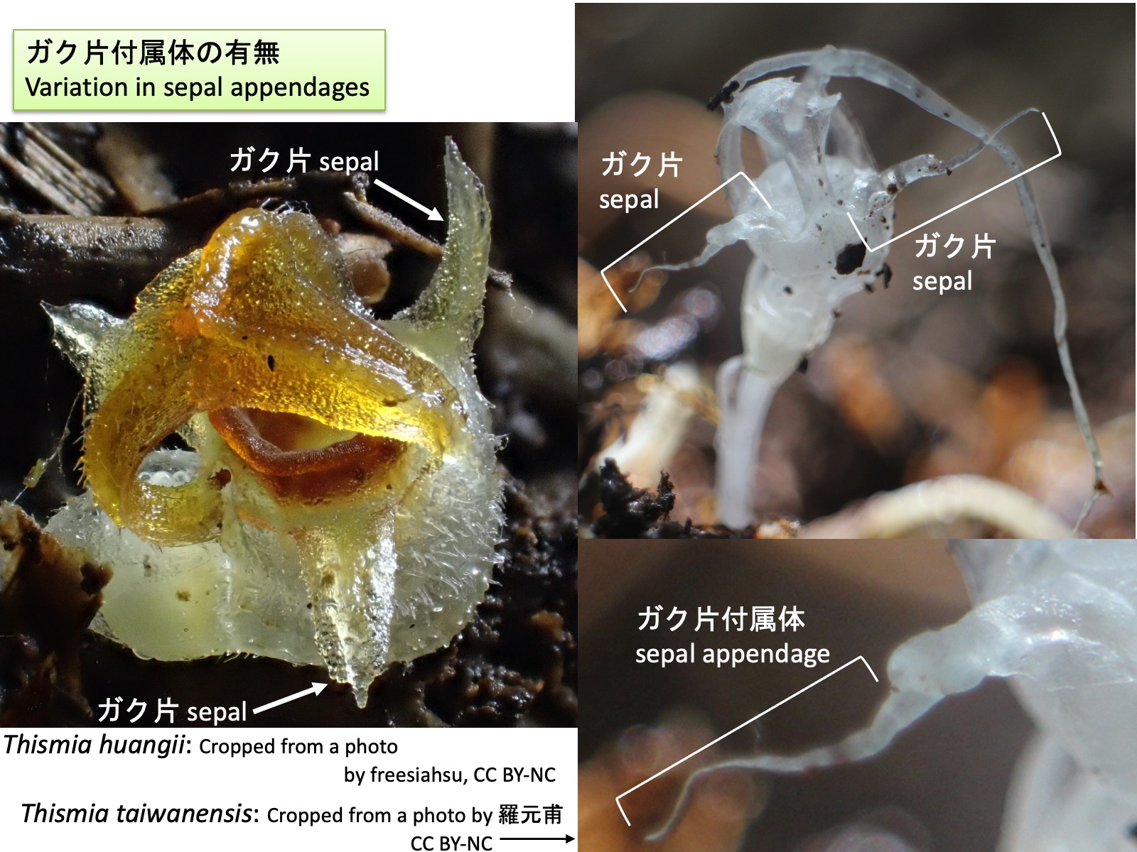
Thismia : Variation in sepal appendages
Thismia taiwanensis: Cropped from a photo by 羅元甫, https://www.inaturalist.org/photos/411535969, CC BY-NC; Thismia huangii: Cropped from a photo by freesiahsu, https://www.inaturalist.org/photos/603575987, CC BY-NC
タヌキノショクダイ属:ガク片付属体の有無
Thismia : Variation in sepal appendages
タヌキノショクダイ属には、ガク片付属体を形成する種としない種がある。Thismia huangii(左写真)ではガク片付属体は形成されないが、T. taiwanensis(右写真)ではガク片先端部が糸状に伸長し、ガク片付属体を形成する。花弁付属体と同様の発生過程を経るのであれば、この付属体はガク片先端部の背軸側に形成された分裂組織に由来する可能性がある。 Some species in Thismia possess sepal appendages, whereas others do not. In Thismia huangii (left photograph), sepal appendages are absent, whereas in T. taiwanensis (right photograph), the distal portion of the sepal elongates into a filamentous sepal appendage. If sepal appendages develop through a process similar to that of petal appendages, they may originate from a meristematic region formed on the abaxial side of the distal portion of the sepal.
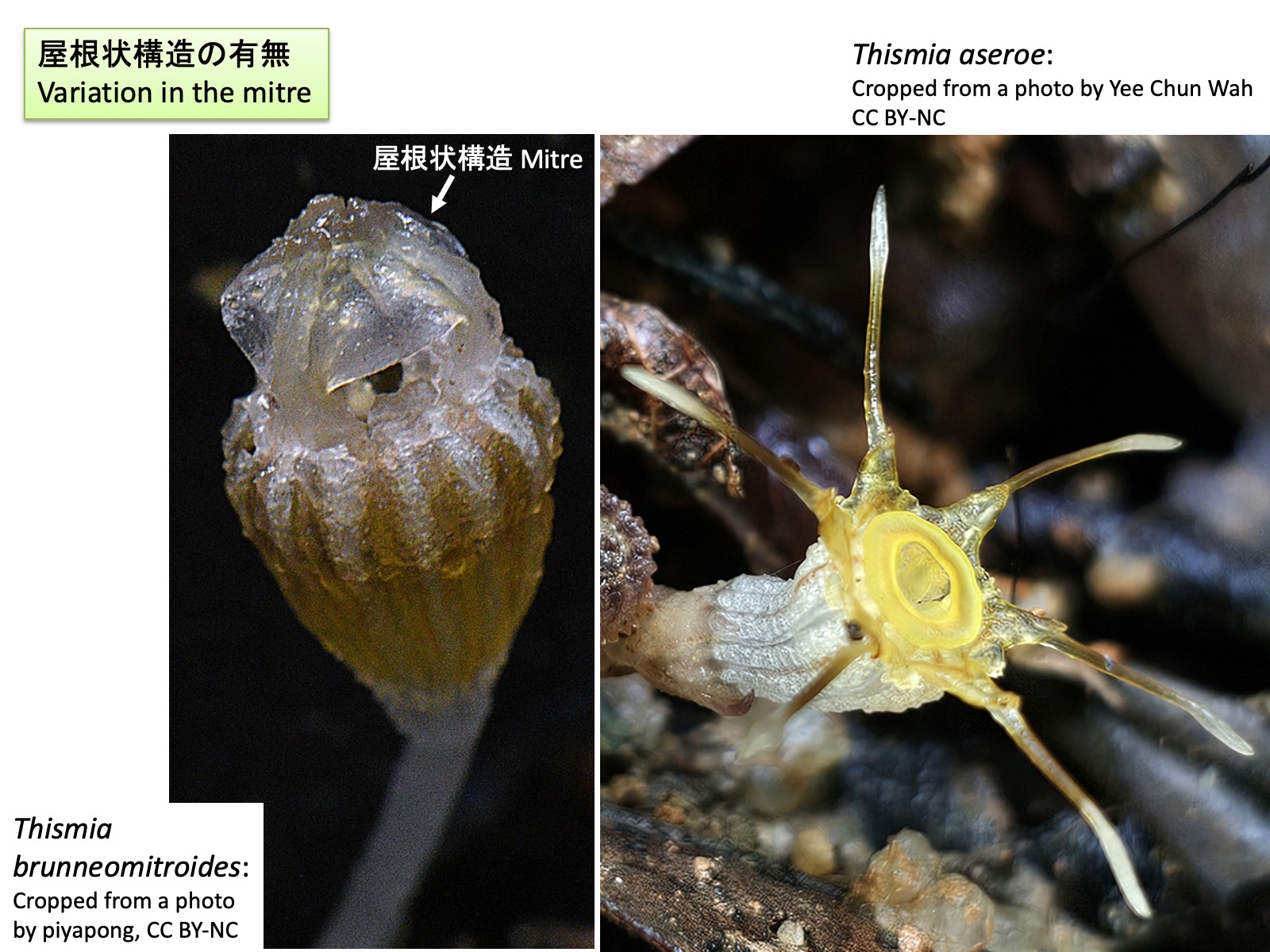
Thismia : Variation in the mitre
Thismia brunneomitroides: Cropped from a photo by piyapong, https://www.inaturalist.org/photos/441605563, CC BY-NC; Thismia aseroe: Cropped from a photo by Yee Chun Wah, https://www.inaturalist.org/photos/588094417, CC BY-NC
タヌキノショクダイ属:屋根状構造の有無
Thismia : Variation in the mitre
タヌキノショクダイ属には、屋根状構造を形成する種と形成しない種がある。Thismia brunneomitroides (左写真)は屋根状構造を形成するのに対し、T. aseroe (右写真)では形成されない。 Within Thismia, some species form a mitre, whereas others do not. Thismia brunneomitroides (left photograph) possesses a mitre, whereas T. aseroe (right photograph) lacks this structure.
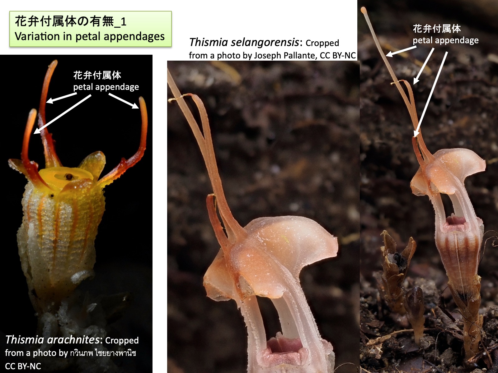
Thismia : Variation in petal appendages_1
Thismia arachnites: Cropped from a photo by กวินภพ ไชยยางพานิช, https://www.inaturalist.org/photos/446284372, CC BY-NC; Thismia selangorensis: Cropped from a photo by Joseph Pallante, https://www.inaturalist.org/photos/443856134, CC BY-NC
タヌキノショクダイ属:花弁付属体の有無_1
Thismia : Variation in petal appendages_1
タヌキノショクダイ属には、花弁付属体を形成する種と形成しない種がある。前図のThismia brunneomitroidesでは花弁付属体は形成されないが、本図のThismia arachnites(左)およびThismia selangorensis(右)では花弁付属体が形成される。 Within Thismia, some species possess petal appendages, whereas others do not. In Thismia brunneomitroides shown in the previous figure, petal appendages are absent, whereas in Thismia arachnites (left) and Thismia selangorensis (right) shown in the present figure, petal appendages are present.
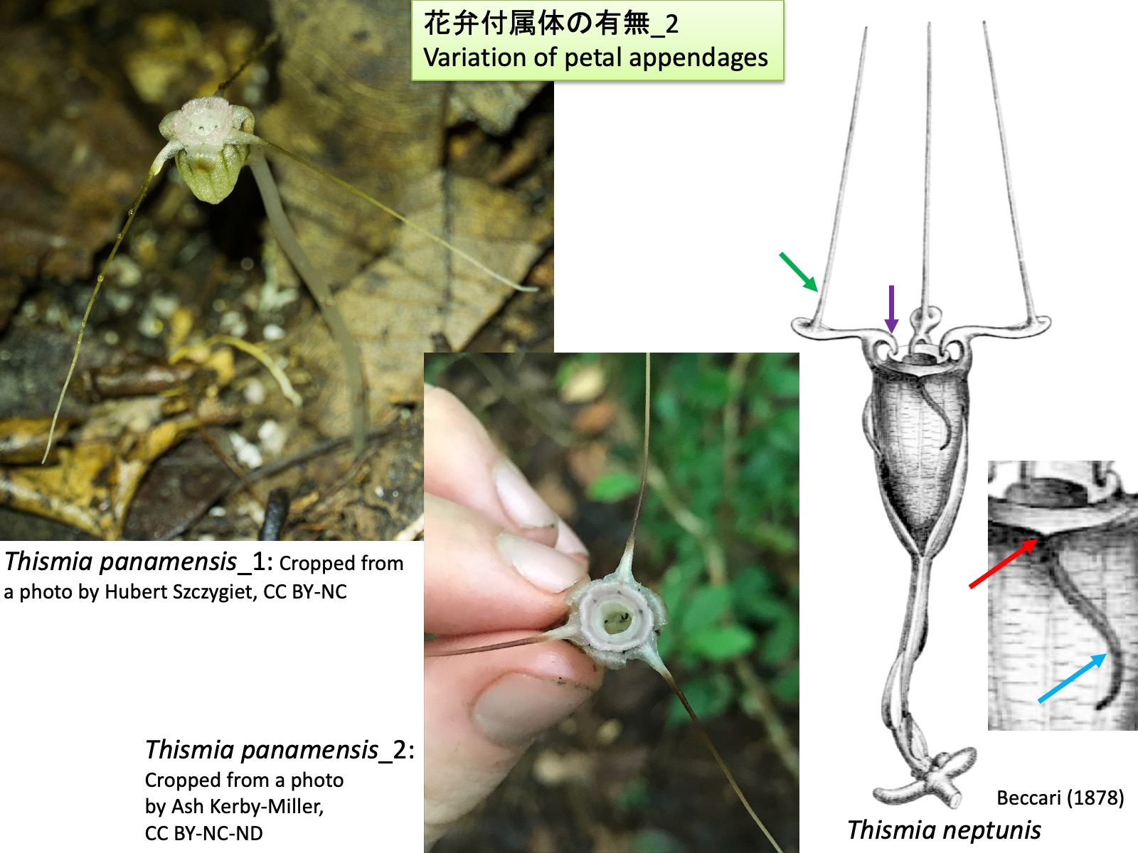
Thismia : Variation in petal appendages_2
Thismia panamensis_1: Cropped from a photo by Hubert Szczygiet, https://www.inaturalist.org/photos/81870711, CC BY-NC; Thismia panamensis_2: Cropped from a photo by Ash Kerby-Miller, https://www.inaturalist.org/photos/58695284, CC BY-NC-ND; Thismia neptunis: Drawings are reproduced from Beccari (1878)
タヌキノショクダイ属:花弁付属体の有無_2
Thismia : Variation in petal appendages_2
分子系統解析 (Shepeleva et al. 2020)において単系統群と推定された旧世界産タヌキノショクダイ属の種では、Nuraliev et al. (2021)が観察したように、花弁の背軸側または向軸側に新たな分裂組織が形成され、それが伸長して花弁付属体や屋根状構造を形成しているように見える。
一方、旧世界産タヌキノショクダイ属とは異なる系統に属し、ティプティニア属Tiputiniaと姉妹群を形成する中南米産のThismia panamensisでは、旧世界産タヌキノショクダイ属の花弁付属体に見られるような、花弁向軸側基部に位置する花弁原基先端と推定される小突起が認められない。このことから、T. panamensisの花弁付属体は、花弁背軸側に異所的な分裂組織が形成されて伸長したものではなく、花弁原基本来の先端部が伸長した構造である可能性がある。なお、ティプティニア属では花弁付属体は形成されない。
さらに、旧世界であるボルネオ島産のThismia neptunisでは、前出のThismia megalongensis と同様に、ガク片付属体が形成される(Sochor et al. 2018)。もし花弁付属体と同様の発生過程を経るのであれば、ガク片向軸側基部付近に見られる突起 (赤色矢印)はガク片原基の先端部に相当し、長い突出部(水色矢印)はガク片原基の背軸側に形成された分裂組織に由来する可能性がある。花弁には、基部側の向軸側に鉤状突起 (紫色矢印)が形成される。この位置は、発生過程が観察された旧世界産タヌキノショクダイ属において屋根状構造を形成する突起(Nuraliev et al. 2021)の位置に類似しているが、形成される突起の形態は大きく異なる。さらに、花弁先端側の向軸側には細長い突起(緑色矢印)が形成される(Sochor et al. 2018)。これは、発生過程が解析された旧世界産タヌキノショクダイ属において、花弁付属体が花弁の背軸側に形成されること(Nuraliev et al. 2021)と対照的である。
以上のことから、旧世界産および新世界産のタヌキノショクダイ属の種には、これまで発生学的に解析された旧世界産タヌキノショクダイ属の突起形成とは異なる形成過程を持つ可能性のある種が存在すると考えられる。今後、発生学的研究と系統解析が進展することで、属内分類の再検討や、異なる種に見られる突起構造の相同性の評価が可能になるであろう。
In the Old World species of Thismia that were inferred to form a monophyletic group in molecular phylogenetic analyses (Shepeleva et al. 2020), petal appendages and mitres appear to be formed through the development of novel meristematic regions on either the abaxial or adaxial side of the petal primordia, followed by their elongation, as observed by Nuraliev et al. (2021).
In contrast, Thismia panamensis, a Neotropical species belonging to a lineage distinct from the Old World species of Thismia and forming a sister group to the genus Tiputinia, lacks the small projection at the adaxial base of the petal that is present in Old World species of Thismia and is thought to represent the original apex of the petal primordium. This observation suggests that the petal appendage of T. panamensis may not originate from an ectopic meristematic region formed on the abaxial side of the petal, but instead may represent an elongation of the original apex of the petal primordium itself. Petal appendages are absent in Tiputinia.
Furthermore, in Thismia neptunis from Borneo in the Old World, sepal appendages are formed (Sochor et al. 2018), as in the previously discussed T. megalongensis. If these structures develop through a process similar to that of petal appendages, the projection located near the adaxial base of the sepal (red arrow) may correspond to the apex of the sepal primordium, whereas the long appendage (light blue arrow) may be derived from a meristematic region formed on the abaxial side of the sepal primordium.
The petals bear hook-shaped projections on their adaxial side near the base (purple arrow). Although these projections occupy a position similar to that of the structures that give rise to the mitre in Old World species of Thismia whose development was studied by Nuraliev et al. (2021), the morphology of the resulting structures differs markedly. In addition, slender projections are formed on the adaxial side near the distal portions of the petals (green arrow) (Sochor et al. 2018). This contrasts with the Old World species of Thismia whose development was analyzed by Nuraliev et al. (2021), in which petal appendages are formed on the abaxial side of the petals. These observations suggest that both Old World and New World species of Thismia may include taxa in which floral appendages are formed through developmental pathways different from those documented in the Old World species studied by Nuraliev et al. (2021). Future developmental and phylogenetic studies will help to reassess infrageneric classification and to evaluate the homology of appendage structures among species.
Pollination mechanisms of Thismia
Thismia tentaculata_1: Cropped from a photo by 桃子, https://www.inaturalist.org/photos/673952674, CC BY-NC; Thismia tentaculata_2: Cropped from a photo by 桃子, https://www.inaturalist.org/photos/673953422, CC BY-NC
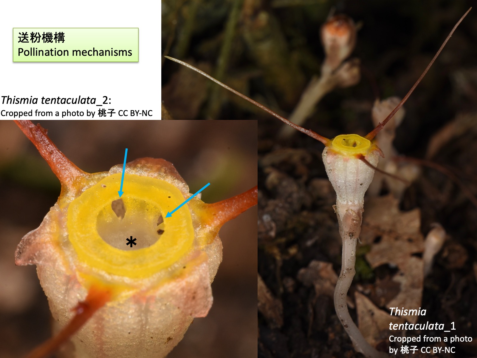
タヌキノショクダイ属の送粉機構
Pollination mechanisms of Thismia
タヌキノショクダイ属の送粉昆虫についてはほとんど研究されていないが、Thismia tentaculataでは詳細な観察が行われている(Guo et al. 2019)。T. tentaculataの花には、キノコバエ類、ショウジョウバエ類、甲虫類、トビムシ類などの訪花が観察されたが、体に花粉を付着させており、有効な送粉者と推定されたのはクロバネキノコバエ科(Sciaridae)の Corynoptera 属のみであった(Guo et al. 2019)。また、日本に自生するタヌキノショクダイにおいても、クロバネキノコバエ科の種が訪花することが報告されている(Suetsugu et al. 2026)。
送粉昆虫はT. tentaculataの黄色の環状部に色で誘引されると考えられており、環状部の開口部(水色矢印)から侵入し、雄ずいと花筒の間の狭い空間を通って花筒基部に位置する雌ずいへと到達する。葯は花筒側に向かって裂開するため、この過程で送粉昆虫の体表に花粉が付着する。雄ずいの葯隔先端部の毛からは液滴が分泌されており、これが送粉昆虫への報酬として機能している可能性も指摘されている。送粉昆虫は花筒底部に到達した後、粘着性の柱頭に接触し、最終的には雄ずいに囲まれた中央の筒状開口部(*)から花外へ脱出する。
タヌキノショクダイ属の花の特異な色彩はキノコへの擬態である可能性が指摘されており、ガク片付属体、花弁付属体、屋根状構造は、キノコバエ類を誘引したり、花上を移動する際の足場として機能しているのではないかと考えられている(Guo et al. 2019)。
Although pollination biology of Thismia has attracted considerable interest, direct observations of pollinators are scarce because the plants are rare, small, and flower on the dark forest floor. Detailed observations have nevertheless been conducted for Thismia tentaculata (Guo et al. 2019). Visitors to the flowers of T. tentaculata included fungus gnats, drosophilid flies, beetles, and springtails. However, the only insects found carrying pollen and therefore considered effective pollinators belonged to the sciarid genus Corynoptera (Sciaridae) (Guo et al. 2019). Similarly, visits by sciarid flies have also been reported in the Japanese species Thismia kobensis (Suetsugu et al. 2026).
Pollinators are thought to be attracted by the yellow annulus of T. tentaculata. They enter the flower through the opening of the annulus (light blue arrows) and move through the narrow space between the stamens and the floral tube to reach the pistil located at the base of the floral tube. Because the anthers dehisce toward the floral tube, pollen is deposited on the body surface of the insect during this process. Droplets are secreted from hairs at the tips of the connectives, and these secretions have been suggested to function as rewards for pollinators. After reaching the base of the floral tube and contacting the sticky stigma, the insects eventually escape from the flower through the central tubular opening (*) surrounded by the stamens. The unusual coloration of Thismia flowers has been suggested to represent mushroom mimicry. The sepal appendages, petal appendages, and mitre may function in attracting fungus gnats and providing footholds for their movement on the flower (Guo et al. 2019).
Haplothismia
Drawings are reproduced from Airy Shaw (1952)
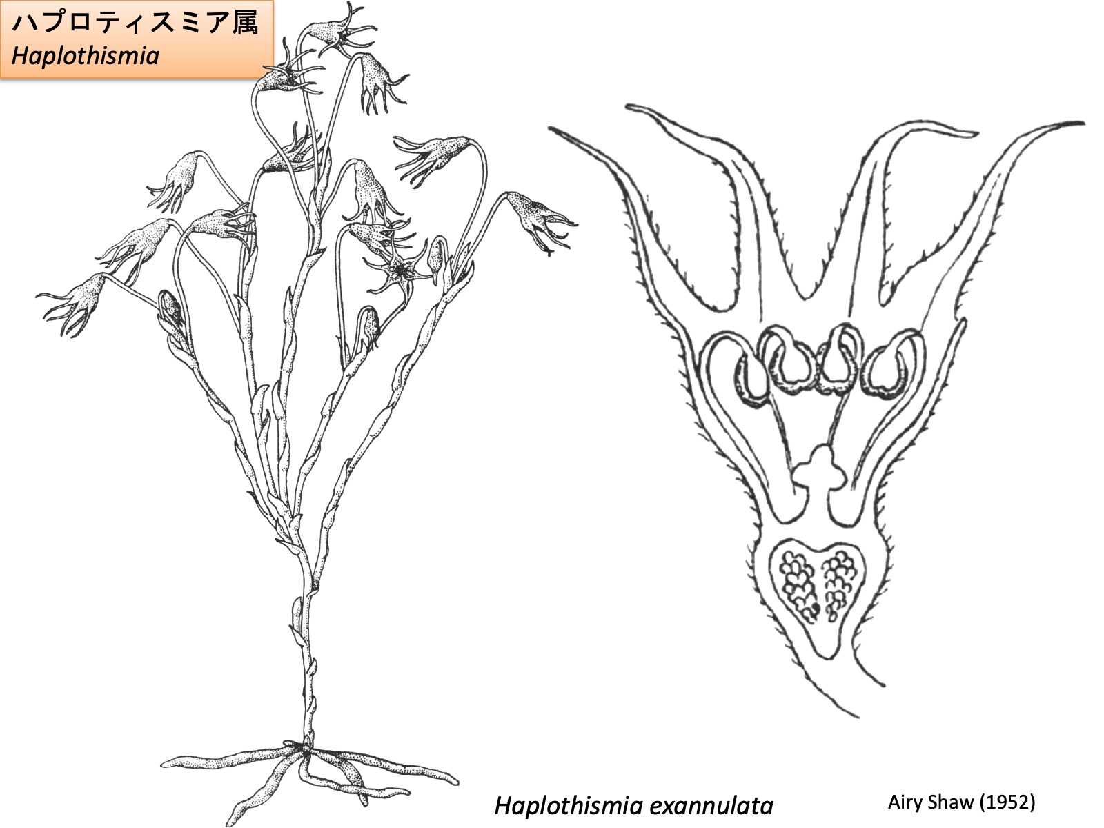
ハプロティスミア属
Haplothismia
ハプロティスミア属の花は、3枚のガク片、3枚の花弁、6本の雄ずい、3心皮が合着した1本の雌ずいからなる (Airy Shaw 1952)。ガク片と花弁は同形であり、その区別は位置に基づく。ハプロティスミア属は、他のタヌキノショクダイ科の属とは異なり、花器官に異所的な突起を形成しない。雄ずいは花筒の外側へ伸長した後、先端部が花筒内側に向かって屈曲すると考えられている。また、花糸基部が異所的に伸長することはなく、環状部も形成されない。
The flower of Haplothismia consists of three sepals, three petals, six stamens, and a single pistil composed of three fused carpels (Airy Shaw 1952). The sepals and petals are morphologically identical and can be distinguished only by their position. Unlike the other genera of Thismiaceae, Haplothismia does not produce ectopic appendages on its floral organs. The stamens are thought to elongate beyond the floral tube and subsequently bend inward toward its interior at their distal ends. In addition, the basal portions of the filaments do not undergo ectopic elongation, and no annulus is formed.

Oxygyne
Oxygyne triandra: Drawings are reproduced from Engler, A. (1908); Oxygyne shinzatoi ホシザキシャクジョウ_1: Cropped from a photo by Hibiki Katayama, https://www.inaturalist.org/photos/328132754, CC BY; Oxygyne shinzatoi ホシザキシャクジョウ_2: Cropped from a photo by Hibiki Katayama, https://www.inaturalist.org/photos/328132800, CC BY; Oxygyne yamashitae ヤクノヒナホシ: Cropped from a photo by Kenji Suetsugu, Thorogood (2019), CC BY-NC-ND 4.0
ヒナノボンボリ属
Oxygyne
ヒナノボンボリ属の花も、3枚のガク片、3枚の花弁、3本の雄ずい、および3心皮が合着した1本の雌ずいからなる (Engler 1908)。ただし、環状部は板状突起によって形成される。Oxygyne triandraの右上図を見ると、この板状突起は花糸基部の向軸側が伸長して形成されたように見える。これは、タヌキノショクダイ属において花糸基部の背軸側が伸長して環状部の隆起を形成するのとは対照的である。さらに、雄ずいは花弁と対生する位置に形成されるが、雄ずいを欠くガク片対生位置にも板状突起が形成される (Yahara and Tsukaya 2008; Suetsugu et al. 2019)。もし板状突起が花糸基部に由来するのであれば、ガク片対生位置においても花糸基部の発生プログラムの一部が保持されている可能性がある。
The flowers of Oxygyne also consist of three sepals, three petals, three stamens, and a single pistil composed of three fused carpels. Unlike those of Thismia, however, the annulus is formed by annulus lamellae. As shown in the upper right figure of Oxygyne triandra, these annulus lamellae appear to be formed by elongation of the adaxial side of the filament base. This contrasts with Thismia, in which the abaxial side of the filament base expands to form the annular ridges. Furthermore, although stamens are produced opposite the petals, lamellar appendages are also present at positions opposite the sepals, where stamens are absent (Yahara and Tsukaya 2008; Suetsugu et al. 2019). If these lamellar appendages are indeed derived from the filament bases, this may indicate that part of the developmental program of the filament base is retained even at the sepal-opposed positions.

ムジナノショクダイ属
Relictithismia
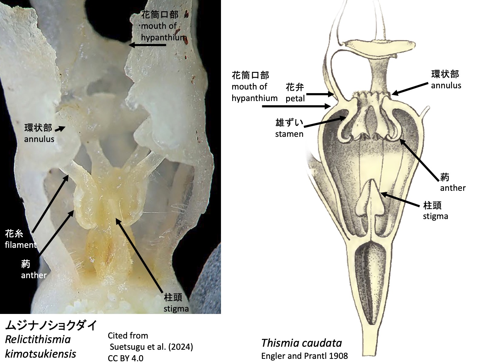
ムジナノショクダイ属の花は、3枚のガク片、3枚の花弁、6本の雄ずい、および3心皮が癒合した1本の雌ずいからなる(Suetsugu et al. 2024)。ガク片と花弁は細長く伸長するが、タヌキノショクダイ属に見られるようなガク片上突起や花弁上突起は見られない。いずれも向軸側基部に機能不明な、丸い細胞からなるカルス状の隆起がある。この隆起は送粉昆虫の誘引に寄与しているのかもしれない。雄ずいは、花筒の口部で花弁から解離するのではなく、口部から全長の1/4ほど基部側の位置で解離する。環状部は雄ずいの解離部付近に形成される。右の写真(Suetsugu et al. 2024)を見ると、雄ずいはタヌキノショクダイ属と同様に、最初から下向きに伸長するように見える。
The flowers of Relictithismia consist of three sepals, three petals, six stamens, and a single pistil formed by the fusion of three carpels (Suetsugu et al. 2024). The sepals and petals are filiform, but they lack the sepal and petal appendages seen in Thismia. Both possess a callus-like cluster composed of rounded cells at the adaxial base, although its function remains unknown. This swelling may contribute to the attraction of pollinating insects. The stamens do not separate from the petals at the mouth of the floral tube, but instead separate at a position about one-quarter of the total length below the mouth. The annulus is formed at the region where the stamens separate. According to the right picture above (Suetsugu et al. 2024), the stamens appear to elongate downward from the outset, as in Thismia.
Tiputinia
Tiputinia foetida_1: Cropped from a photo by Alan Rockefeller, https://www.inaturalist.org/photos/362476435, CC BY; Tiputinia foetida_2: Cropped from a photo by Alan Rockefeller, https://www.inaturalist.org/photos/366763162, CC BY; Tiputinia foetida_3: Cropped from a photo by Alan Rockefeller, https://www.inaturalist.org/photos/367555871, CC BY
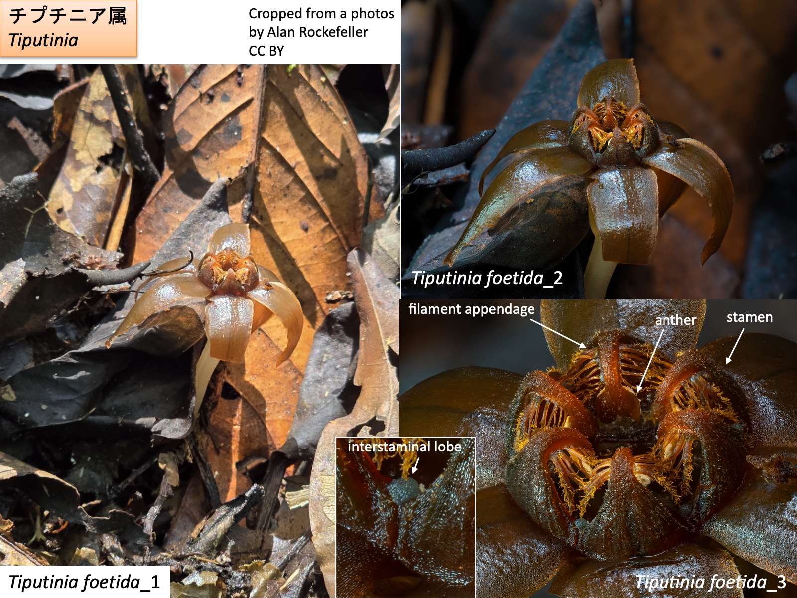
チプチニア属
Tiputinia
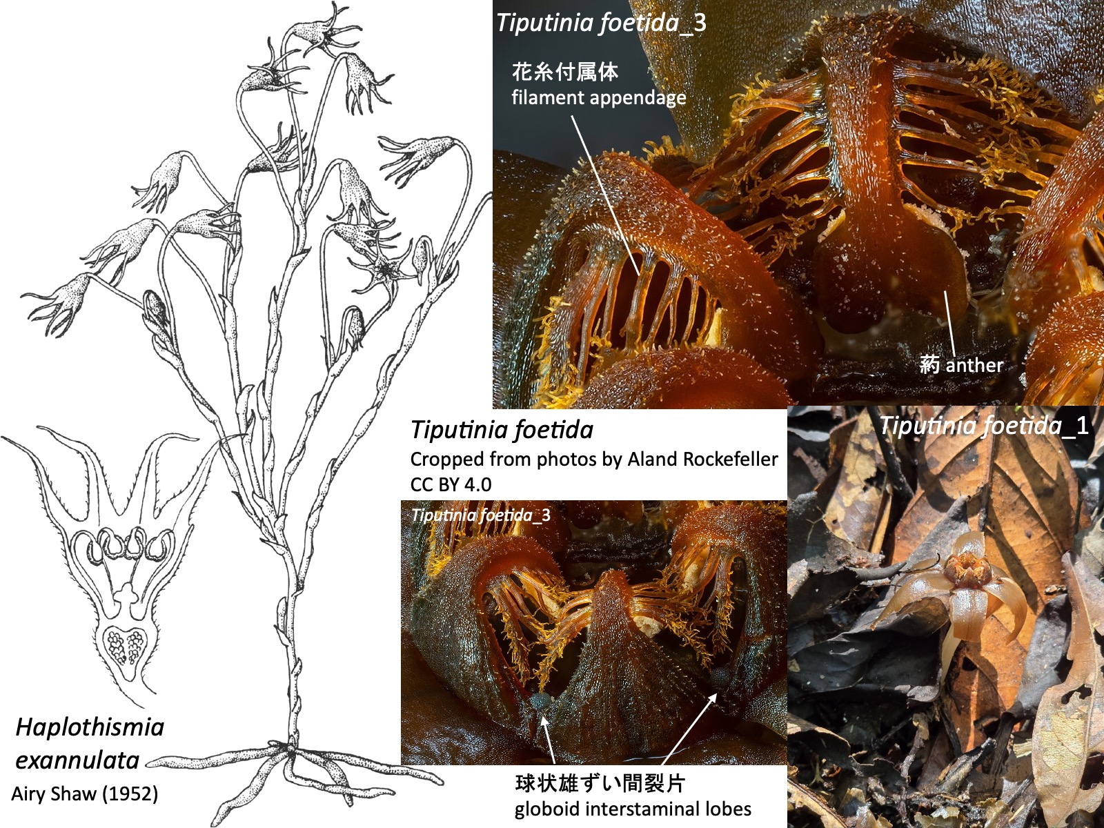
チプチニア属の花は、3枚のガク片、3枚の花弁、6本の雄ずい、および3心皮が癒合した1本の雌ずいからなる (Woodward et al. 2007)。雄ずいは花筒の外側へ伸び、途中で花筒の内側へ向けて屈曲する。花糸基部は広がって環状につながるが、花筒内側へ張り出す環状部annulusは形成されない。花糸からは花糸付属体が伸長し、雄ずい間には球状の雄ずい間裂片 globoid interstaminal lobesが形成される。
The flowers of Tiputinia consist of three sepals, three petals, six stamens, and a single pistil formed by the fusion of three carpels (Woodward et al. 2007). The stamens extend outward from the floral tube and then bend inward toward the inside of the tube. The filament bases are expanded and connected in a ring, but they do not form an annulus projecting into the floral tube. Filament appendages extend from the filaments, and globoid interstaminal lobes are formed between the stamens.
タヌキノショクダイ科の属の検索表(Cheek et al. 2018とSuetsugu et al. 2024を参考に作成)
Key to the genera of Thismiaceae (based on Cheek et al. 2018 and Suetsugu et al. 2024)
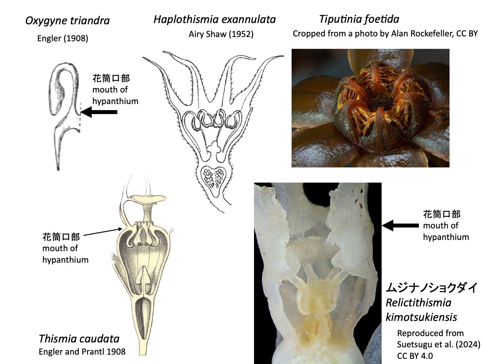
1a. 雄ずいの花糸は花筒の口部で花弁と離生する。雄ずいははじめは花筒外部へ伸長するが、後に花筒内側へ屈曲する。屈曲部は花筒口部外側から見える。葯は花筒口部か、花筒のやや内部に位置する。…… 2 (Oxygyne ヒナノボンボリ属, Haplothismia ハプロティスミア属、Tiputinia ティプティニア属)
1b. 雄ずいの花糸は花筒内部(Relictithismia ムジナノショクダイ属)または花筒口部(Thismia タヌキノショクダイ属)で花弁と離生する。葯は花筒内部に位置する。 …… 4(Relictithismia ムジナノショクダイ属、Thismia タヌキノショクダイ属)
1a. Staminal filaments free from the perianth at the mouth of the hypanthium. Stamens initially extend outward from the hypanthium but later bend inward; the bent portion is visible from outside the mouth of the hypanthium. Anthers positioned at the mouth of the hypanthium or slightly within it. …… 2 (Oxygyne, Haplothismia, Tiputinia)
1b. Staminal filaments free from the perianth within the hypanthium (in Relictithismia) or at the mouth of the hypanthium (in Thismia). Anthers positioned within the hypanthium. …… 4 (Relictithismia, Thismia)
2a. 雄ずいは6本。環状部(annulus)を形成しない。 …… 3(Haplothismia ハプロティスミア属、Tiputinia ティプティニア属)
2b. 雄ずいは3本。環状部に板状突起(annulus lamellae)を形成する。 …… Oxygyne ヒナノボンボリ属
2a. Stamens 6. Annulus absent. …… 3 (Haplothismia, Tiputinia)
2b. Stamens 3. Annulus present and bearing lamellar appendages (annulus lamellae). …… Oxygyne
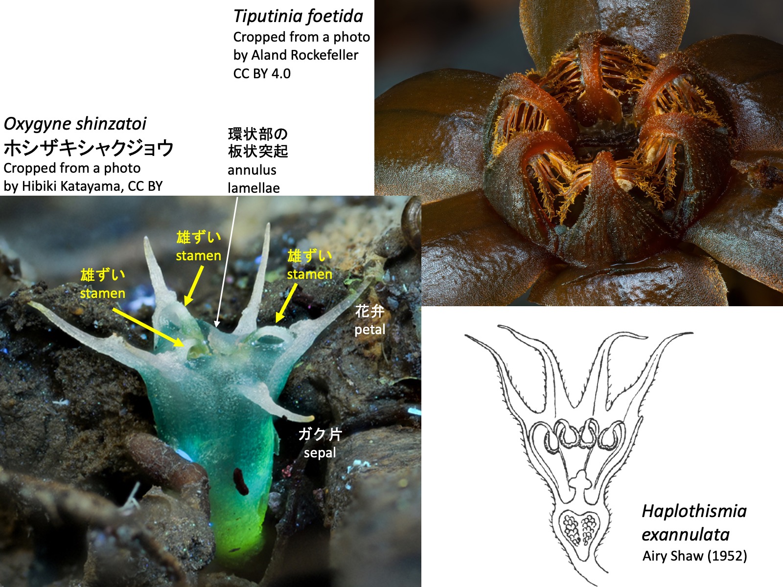
3a. 花序は地上性で、多数回分枝し、各枝の先端に花をつける。雄ずい上に異所的な突起は形成されない。 …… Haplothismia ハプロティスミア属
3b. 花序は地表近くに生じ、通常は1花のみをつける。雄ずいの花糸から5対または6対の花糸付属体 filament appendageを形成する。球状の雄ずい間裂片(globoid interstaminal lobes)を形成する。 …… Tiputinia ティプティニア属
3a. Inflorescence aerial, repeatedly branched, with flowers borne at the tips of the branches. Stamens lacking ectopic appendages. …… Haplothismia
3b. Inflorescence produced close to the ground surface, usually bearing a single flower. Stamens with five or six pairs of filament appendages arising from the filaments. Globoid interstaminal lobes present. …… Tiputinia
Tiputinia foetida_3: Cropped from a photo by Alan Rockefeller, https://www.inaturalist.org/photos/367555871, CC BY; Haplothismia exannulata: Reproduced from Airy Shaw (1952)
4a. 雄ずいの花糸は花筒内部で花弁と離生し、環状部は花筒内部に形成される。葯背側が柱頭に付着する。 …… Relictithismia ムジナノショクダイ属
4b. 雄ずいの花糸は花筒口部で花弁と離生し、環状部は花筒口部に形成される。葯は柱頭と離れて位置する。 …… Thismia タヌキノショクダイ属
4a. Staminal filaments free from the perianth within the hypanthium, and the annulus formed within the hypanthium. The abaxial side of the anthers adheres to the stigma. …… Relictithismia
4b. Staminal filaments free from the perianth at the mouth of the hypanthium, and the annulus formed at the mouth of the hypanthium. Anthers separated from the stigma. …… Thismia
謝辞
Acknowledgements
タヌキノショクダイ科をまとめるにあたり、塚谷裕一博士、末次健司博士、滝澤和馬氏、乾和輝氏にはたいへんお世話になりました。心より御礼申し上げます。ただし、本稿に記載した内容に関する責任は長谷部光泰にあります。
I am deeply grateful to Dr. Hirokazu Tsukaya, Dr. Kenji Suetsugu, Mr. Kazuma Takizawa, and Mr. Kazuki Inui for their invaluable help in preparing this account of Thismiaceae. Responsibility for all contents of this manuscript, however, rests solely with Mitsuyasu Hasebe.
引用文献
References
Airy Shaw, H.K. (1952). A new genus and species of Burmanniaceae from South India. Kew Bull. 7, 277. https://doi.org/10.2307/4109280.
Beccari, O. (1878) Burmanniaceae. Malesia 1: 240-253.
Cheek, M., Tsukaya, H., Rudall, P.J., and Suetsugu, K. (2018). Taxonomic monograph of Oxygyne (Thismiaceae), rare achlorophyllous mycoheterotrophs with strongly disjunct distribution. PeerJ : e4828. https://doi.org/10.7717/PEERJ.4828/FIG-10.
Engler, A. (1889) Burmanniaceae. In Engler, A. and Prantl, K. Die Natürlichen Pflanzen Familien. II Teil. 6. Abteilung. page 47, Fig. 38. Leipzig, Verlag von Wilhelm Engelmann.
Engler, A. (1908) Die Pflanzenwelt Afrikas insbesondere seiner tropischen Gebiete. Vol. 2 page 9. In: Engler & Drude (eds.), Die Vegetation der Erde 9. Leipzig: Wilhelm Engelmann.
Engler, A. and Prantl, K. 1908. Nat. Pflanzenfam. Erganzungshefte II. p. 73 Fig. 12. Thismia caudataがシノニムのGlaziocharis macahensisとして図示されている。Thismia caudata is illustrated under its synonym Glaziocharis macahensis.
Schlechter, R. (1919) 57. Eine neue Papuasische Burmanniacee. In Engler, A. (1919) Bot. Jahrb. Syst. 55: 202-203
Feller, B., Dančák, M., Hroneš, M., Sochor, M., Suetsugu, K., and Imhof, S. (2022). Mycorrhizal structures in mycoheterotrophic Thismia spp. (Thismiaceae): functional and evolutionary interpretations. Mycorrhiza 32, 269–280. https://doi.org/10.1007/S00572-022-01076-3.
Gomes, S.I.F., Aguirre-Gutiérrez, J., Bidartondo, M.I., and Merckx, V.S.F.T. (2017). Arbuscular mycorrhizal interactions of mycoheterotrophic Thismia are more specialized than in autotrophic plants. New Phytol. 213, 1418–1427. https://doi.org/10.1111/NPH.14249;WGROUP:STRING:PUBLICATION.
Guo, X., Zhao, Z., Mar, S.S., Zhang, D., and Saunders, R.M.K. (2019). A symbiotic balancing act: arbuscular mycorrhizal specificity and specialist fungus gnat pollination in the mycoheterotrophic genus Thismia (Thismiaceae). Ann. Bot. 124, 331–342. https://doi.org/10.1093/AOB/MCZ087.
Kumar, P., Gale, S.W., Li, J.-H., Bouamanivong, S., and Fischer, G.A. (2017). Thismia nigricoronata, a new species of Burmanniaceae (Thismieae, Dioscoreales) from Vang Vieng, Vientiane Province, Laos, and a key to subgeneric classification. Phytotaxa 319, 225–240. https://doi.org/10.11646/PHYTOTAXA.319.3.2.
Merckx, V.S.F.T., Gomes, S.I.F., Wapstra, M., Hunt, C., Steenbeeke, G., Mennes, C.B., Walsh, N., Smissen, R., Hsieh, T.H., Smets, E.F., et al. (2017). The biogeographical history of the interaction between mycoheterotrophic Thismia (Thismiaceae) plants and mycorrhizal Rhizophagus (Glomeraceae) fungi. J. Biogeogr. 44, 1869–1879. https://doi.org/10.1111/JBI.12994;PAGE:STRING:ARTICLE/CHAPTER.
Nuraliev, M.S., Yudina, S. V., Shepeleva, E.A., Truong, B.V., Do, T.X., Beer, A.S., and Remizowa, M. V. (2021). Floral structure in Thismia (Thismiaceae: Dioscoreales): new insights from anatomy, vasculature and development. Bot. J. Linn. Soc. 195, 501–531. https://doi.org/10.1093/BOTLINNEAN/BOAA066.
Parniske, M. (2008). Arbuscular mycorrhiza: the mother of plant root endosymbioses. Nat. Rev. Microbiol. 6, 10 6, 763–775. https://doi.org/10.1038/nrmicro1987.
POWO (2026). Plants of the World Online. Facilitated by the Royal Botanic Gardens, Kew. Published on the Internet; https://powo.science.kew.org/. Retrieved 3 June 2026. Distribution data from Kew Backbone Distributions (WCVP). © World Checklist of Vascular Plants, licensed under CC BY 3.0.
Ríos, R.C., and da Cruz, V.J.M.V. (2023). Rediscovery of Thismia caudata after 129 years and synopsis of other achlorophyllous herbs from the Pico do Marumbi State Park and buffer zone in Piraquara, Paraná, Brazil. Rodriguésia 74, e01122022. https://doi.org/10.1590/2175-7860202374042.
Shepeleva, E.A., Schelkunov, M.I., Hroneš, M., Sochor, M., Dančák, M., Merckx, V.S., Kikuchi, I.A., Chantanaorrapint, S., Suetsugu, K., Tsukaya, H., et al. (2020). Phylogenetics of the mycoheterotrophic genus Thismia (Thismiaceae: Dioscoreales) with a focus on the Old World taxa: delineation of novel natural groups and insights into the evolution of morphological traits. Bot. J. Linn. Soc. 193: 287–315. https://doi.org/10.1093/BOTLINNEAN/BOAA017.
Sochor, M., Egertová, Z., Hroneš, M., and Dančák, M. (2018). Rediscovery of Thismia neptunis (Thismiaceae) after 151 years. Phytotaxa 340, 71–78. https://doi.org/10.11646/PHYTOTAXA.340.1.5.
末次健司(2023)「植物」をやめた植物たち(福音館書店)
Suetsugu, K., Sugimoto, T., and Tsukaya, H. (2019). Emended description and new localities of Oxygyne shinzatoi. (Burmanniaceae/Thismiaceae), with discussion of phylogenetic relationships of Oxygyne from Japan and Africa. Phytotaxa 423, 238-246. https://doi.org/10.11646/PHYTOTAXA.423.4.2.
Suetsugu, K., Nakamura, Y., Nakano, T., and Tagane, S. (2024). Relictithismia kimotsukiensis, a new genus and species of Thismiaceae from southern Japan with discussions on its phylogenetic relationship. J. Plant Res. 137: 411–422. https://doi.org/10.1007/S10265-024-01532-5.
Suetsugu, K., Sennikov, A.N., and Nuraliev, M.S. (2026). Multifaceted obscurity of Thismia abei (Thismiaceae): A fairy lantern with the protologue long disregarded in practice. Plants People Planet. https://doi.org/10.1002/PPP3.70214;JOURNAL:JOURNAL:25722611;REQUESTEDJOURNAL:JOURNAL:25722611;WGROUP:STRING:PUBLICATION.
Thorogood, C.J. (2019). Oxygyne: An extraordinarily elusive flower. Plants People Planet 1, 67–70. https://doi.org/10.1002/PPP3.26;JOURNAL:JOURNAL:25722611;REQUESTEDJOURNAL:JOURNAL:25722611;WGROUP:STRING:PUBLICATION.
塚谷裕一 (2016) 森を食べる植物 (岩波書店)
Woodward, C.L., Berry, P.E., Kamer, H.M. de, and Swing, K. (2007). Tiputinia foetida, a new mycoheterotrophic genus of Thismiaceae from Amazonian Ecuador, and a likely case of deceit pollination. Taxon 56, 157–162. https://doi.org/10.2307/25065746. Yahara, T., and Tsukaya, H. (2008). Oxygyne yamashitae, a new species of Thismiaceae from Yaku island, Japan. Acta Phytotax. Geobot. 59, 97–104. https://doi.org/10.18942/APG.KJ00005012322.