National Institute for Basic Biology


2013.02.18
The research group of Associate Professor Shigenori Nonaka and researcher
Daisuke Takao, Laboratory for Spatiotemporal Regulations at NIBB, have found out an asymmetric distribution of dynamic calcium signals in the node of mouse embryo during left-right axis formation.
In the node of the mouse embryo, rotational movements of cilia generate an external liquid flow known as nodal flow, which determines left-right asymmetric gene expression. How nodal flow is converted into asymmetric gene expression is still controversial, but the increase of Ca2+ levels in endodermal cells to the left of the node has been proposed to play a role.However, Ca2+ signals inside the node itself have not yet been described.
Using an optimized Ca2+ imaging method, the group was able to observe dynamic Ca2+ signals in the node in live mouse embryos. Pharmacological disruption of Ca2+ signals did not affect ciliary movements or nodal flow, but did alter the expression patterns of the Nodal and Cerl-2 genes. Quantitative analyses of Ca2+ signal frequencies and distributions showed that during left-right axis establishment, formerly symmetric Ca2+ signals became biased to the left side. In iv/iv mutant embryos that showed randomized laterality due to ciliary immotility, Ca2+ signals were found to be variously left-sided, right-sided, or bilateral, and thus symmetric on average. In Pkd2 mutant embryos, which lacked polycystin-2, a Ca2+-permeable cation channel necessary for left-right axis formation, the Ca2+ signal frequency was lower than in wild-type embryos. These data support a model in which dynamic Ca2+ signals in the node are involved in left-right patterning.

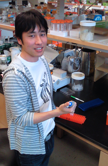
Dr. Daisuke Takao