<?xml version="1.0" encoding="UTF-8"?>
<feed xmlns="http://www.w3.org/2005/Atom">
<title>NIBB / News</title>
    <link rel="alternate" type="text/html" href="https://www.nibb.ac.jp/en/pressroom/" />
    <link rel="self" type="application/atom+xml" href="" />
    <id>tag:www.nibb.ac.jp,2011-06-10:/pressroom//6</id>
    <updated>2026-03-04T04:58:52Z</updated>


<entry>
<title>[Press Release] Rethinking How Nucleoside Supplementation Speed Up DNA Replication: It’s Not Just About Having Enough</title>
    <link rel="alternate" type="text/html" href="https://www.nibb.ac.jp/en/pressroom/news/2025/10/24.html" />
    <id>tag:www.nibb.ac.jp,2025:/pressroom/news//7.10392</id>

    <published>2025-10-24T01:00:00Z</published>
    <updated>2025-10-24T02:19:48Z</updated>

<author>
        <name>NIBB - OFFICE OF PUBLIC RELATIONS AND INTERNATIONAL COOPERATION</name>
    </author>
    
        <category term="Press Release"/>
    
    
    <content type="html" xml:lang="en" xml:base="https://www.nibb.ac.jp/pressroom/news/">
        <![CDATA[DNA, often called the blueprint of life, is built from four chemical building blocks known as deoxy-nucleoside triphosphates (dNTPs)&mdash;dATP, dTTP, dGTP, and dCTP. Cells constantly need these molecules to duplicate their DNA during active proliferation. When the supply runs low, DNA replication slows or stops, halting cell proliferation.<br />
<br />
To prevent this, researchers often add nucleosides(A, T, G, and C)&mdash;the precursors of dNTPs&mdash;to the cell culture medium. This treatment is known to help DNA replication proceed smoothly and has long been assumed to work simply by increasing the overall pool of dNTPs inside the cell. However, the exact mechanism behind this effect has remained unclear.<br />
A research team led by Andrei Chabes (Ume&aring; University, Sweden), in collaboration with labs of Tomomi Tsubouchi (National Institute for Basic Biology/Shizuoka University, Japan) and Erik Johansson (Ume&aring; University, Sweden) has now uncovered a surprising answer. In their recent paper in Nucleic Acids Research, Pandey et al. report that the key effect of nucleoside supplementation is not a general boost in all DNA components&mdash;but a specific balancing act involving T (thymidine).<br />
<br />
The researchers found that adding T helps cells counteract an inhibitory molecule, dUTP, a faulty DNA building block that can perturb DNA synthesis. Supplying T increases the correct molecule, dTTP, which prevents dUTP from interfering with DNA replication.<br />
&ldquo;We have routinely used nucleosides as a tool to accelerate replication, but we were uncertain what was really happening inside the cell,&rdquo; said Dr. Kiminori Kurashima, one of the study&rsquo;s authors. &ldquo;It was satisfying to finally see the quantitative effects&mdash;and the results clearly showed that the long-held assumption was incorrect.&rdquo;<br />
<br />
To further test this idea, the team increased dUTP levels in cells and observed that DNA replication slowed down. Adding T restored normal replication speed. Even in a purified test-tube system, they confirmed that dUTP directly inhibits the enzyme responsible for copying DNA.<br />
<br />
&quot;For many years, the question of how nucleosides accelerate DNA replication remained out of reach simply because measuring dNTPs precisely is so difficult,&rdquo; said Tomomi Tsubouchi. &ldquo;Thanks to Praveen Pandey&rsquo;s precise dNTP measurements in the Chabes laboratory and the Johansson lab&rsquo;s reconstitution of DNA synthesis&nbsp;<em>in vitro</em>, we were able to see how nucleoside supplementation can accelerate replication.<br />
<br />
This study refines the long-standing view that nucleosides simply promote DNA replication by increasing the total pool of DNA building blocks. It shows that elevated dTTP&mdash;by limiting dUTP&rsquo;s inhibitory effect at DNA polymerase&mdash;also plays a critical role in smooth, accurate replication.<br />
<br />
＿＿＿<br />
<br />
<div>
	<em>Nucleic Acids Research</em></div>
<div>
	Decoding Nucleoside Supplementation: How Thymidine Outperforms Ribonucleosides in Accelerating Mammalian Replication Forks</div>
<div>
	Praveen Pandey, Kiminori Kurashima, G&ouml;ran Bylund, Erik Johansson, Tomomi Tsubouchi and Andrei Chabes</div>
<div>
	DOI:&nbsp;<a href="https://doi.org/10.1093/nar/gkaf1035">https://doi.org/10.1093/nar/gkaf1035</a></div>
<div>
	&nbsp;</div>
]]>
    </content>
</entry>


<entry>
<title>[Press Release] Discovery of a Novel Intracellular Trafficking Pathway in Plant Cells</title>
    <link rel="alternate" type="text/html" href="https://www.nibb.ac.jp/en/pressroom/news/2025/10/03.html" />
    <id>tag:www.nibb.ac.jp,2025:/pressroom/news//7.10382</id>

    <published>2025-10-03T09:00:00Z</published>
    <updated>2025-10-03T17:10:48Z</updated>

<author>
        <name>NIBB - OFFICE OF PUBLIC RELATIONS AND INTERNATIONAL COOPERATION</name>
    </author>
    
        <category term="Press Release"/>
    
    
    <content type="html" xml:lang="en" xml:base="https://www.nibb.ac.jp/pressroom/news/">
        <![CDATA[Just like in yeast and animal cells, vacuoles in plants are responsible for breaking down unwanted cellular components. At the same time, vacuoles in seeds also serve the opposite role -storing large amounts of proteins that provide energy during germination. These storage proteins, accumulated in the vacuoles of seeds such as beans and wheat, are not only vital for plant growth but also represent an important agricultural resource closely tied to our daily diet.<br />
<br />
Until now, it was entirely unknown whether proteins could be transported&nbsp;<em>from</em>&nbsp;the vacuole to other organelles. In a study now published in&nbsp;<em>Nature Plants</em>, Dr. Yihong Feng (Specially Appointed Assistant Professor) and Professor Takashi Ueda at the National Institute for Basic Biology in Japan, together with their collaborators, demonstrated in the model plant&nbsp;<em>Arabidopsis thaliana</em>&nbsp;the existence of a retrograde trafficking pathway that retrieves the membrane protein VAMP727 from the vacuolar membrane back to endosomes. The team further identified the molecular machinery responsible for this pathway.<br />
Dr. Feng commented, &ldquo;The sorting nexin proteins that function in this newly discovered pathway have independently diversified in plants compared to animals and yeast. Our findings indicate that this trafficking route is a plant-specific innovation.&rdquo;<br />
Professor Ueda added, &ldquo;We were able to show that the plant-unique membrane fusion protein VAMP727 co-evolved with a recycling mechanism from the vacuole. This suggests that the evolution of vacuolar protein transport in seed plants, which is essential for massive storage protein accumulation, was closely tied to the emergence of this retrograde pathway.&rdquo;<br />
<br />
This study reveals part of the unique membrane trafficking network that plants have evolved during their history, providing new insights into both cell biology and plant science.<br />
&nbsp;<br />
<img alt="efig1.jpg" class="mt-image-center" src="https://www.nibb.ac.jp/pressroom/news/uploads/20251003/efig1.jpg" style="text-align: center; display: block; margin: 0px auto 20px;" /><strong>
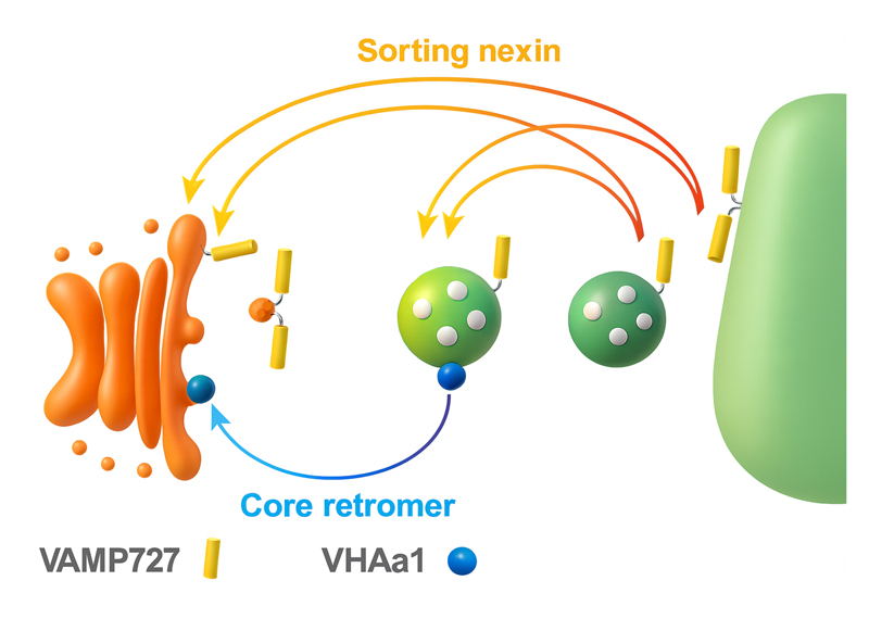
Figure 1. Retrograde trafficking pathways from the plant vacuole and endosomes.</strong><br />
This study revealed two independent retrograde pathways: a&nbsp;sorting nexin&ndash;mediated pathway&nbsp;(orange) and a&nbsp;core retromer&ndash;mediated pathway&nbsp;(blue). In yeast, sorting nexins and the core retromer form a single complex that functions in endosome-to-Golgi retrograde transport. In contrast, our findings demonstrate that in plants, SNX and the core retromer operate independently, mediating distinct retrograde routes with different cargo proteins. Together, these pathways establish and maintain the unique membrane protein compositions of vacuoles and endosomes.<br />
&nbsp;<br />
<img alt="efig2.jpg" class="mt-image-center" src="https://www.nibb.ac.jp/pressroom/news/uploads/20251003/efig2.jpg" style="text-align: center; display: block; margin: 0px auto 20px;" /><strong>
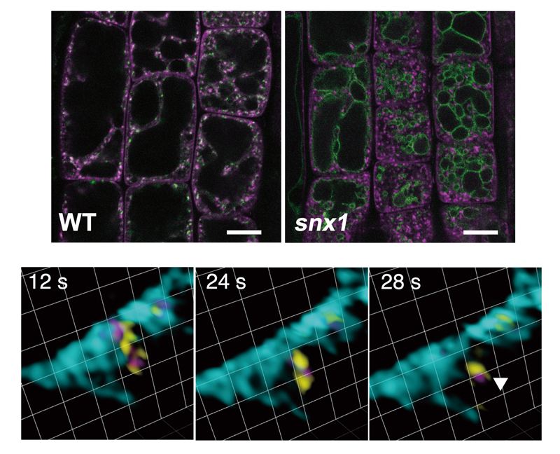
Figure 2. VAMP727 is retrieved from the vacuolar membrane by SORTING NEXINs.</strong><br />
<strong>Top panels:</strong>&nbsp;Root epidermal cells expressing Venus-VAMP727 and mRFP-VAMP721 in wild type (left) and the <em>snx1</em> mutant (right). The punctate localization pattern of VAMP721, which functions in the secretory pathway, remained unchanged in the <em>snx1</em> mutant. In contrast, VAMP727 accumulated at the vacuolar membrane in the <em>snx1</em> background. Scale bar = 10 &micro;m.<br />
<strong>Bottom panels:</strong>&nbsp;Images acquired using the super-resolution confocal live imaging microscope (SCLIM) of mGFP-VAMP727 (yellow), SNX1-mRFP (magenta), and the vacuolar membrane marker miRFP-SYP22 (cyan). These images capture the budding of VAMP727 together with SNX1 from the vacuolar membrane. Grid size = 0.942 &micro;m.<br />
&nbsp;<br />
<img alt="efig3.jpg" class="mt-image-center" src="https://www.nibb.ac.jp/pressroom/news/uploads/20251003/efig3.jpg" style="text-align: center; display: block; margin: 0px auto 20px;" /><strong>
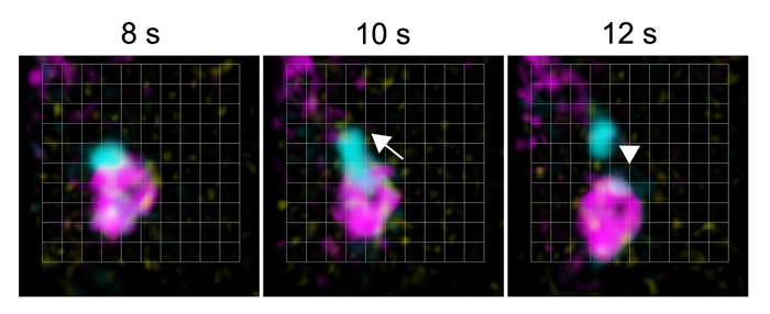
Figure 3. SNX and the core retromer function in distinct retrograde pathways.</strong><br />
Images acquired using the SCLIM of VPS35b-iRFP (core retromer, cyan), SNX1-mRFP (magenta), and the endosomal marker GFP-ARA7 (yellow). Time-lapse observations revealed budding of VPS35b from the endosomal membrane, which did not include SNX1. Grid size = 0.426 &micro;m.<br />
<br />
<br />
<em>Nature Plants</em><br />
Retrieval from vacuolar/endosomal compartments underpinning neofunctionalization of SNARE in plants<br />
Yihong Feng, Kazuo Ebine, Yoko Ito, Takehiko Kanazawa, Tatsuya Sawasaki, Akira Nozawa, Tomohiro Uemura, Akihiko Nakano, Takashi Ueda<br />
DOI： <a href="https://doi.org/10.1038/s41477-025-02115-5">10.1038/s41477-025-02115-5</a><br />
<br />
]]>
    </content>
</entry>


</feed>
