日本語

National Institutes of Natural Sciences

National Institute for Basic Biology

NIBB Departments

Researcher Infomation

OHTA, Yusaku
Specially Appointed Assistant Professor
OHTA, Yusaku
Affiliation:

Research Summary

Image analysis is an important component of understanding life sciences. It enables us to quantify phenomena by extracting meaningful information from a large number of images and to properly represent that information. I am developing image analysis techniques aimed at analyzing the developmental process of early zebrafish embryo at whole embryo scale and single-cell resolution. Currently, I am developing an analysis technique that can simultaneously extract multiple pieces of information such as cell morphology, cell motility, and cell dynamics by combining 3D cell tracking and functional imaging.

I. Simultaneous multifunctional analysis of early embryonic development at a whole embryo scale and single cell resolution.

Three-dimensional remodeling of cell populations through cell migration is essential in early embryogenesis. Cell migration is highly coordinated by controlling cell-cell adhesion. To elucidate the principles of such complex embryogenesis, it is necessary to understand the cellular dynamics of the whole embryo at single cell resolution. I am developing image analysis techniques to analyze the developmental process of early zebrafish embryogenesis at whole-embryo scale and single-cell resolution.
 
With the development of microscopy, imaging of early embryonic imaging has evolved from two-dimensional, embryonic development has evolved from two-dimensional, fixed specimen and partial embryo observation to three-dimensional, living specimen and whole embryo scale. This evolution has led to an explosion in the analysis of cell migration during the development of early embryos in recent years. However, conventional image analysis techniques could only extract information on cell migration in early development. Therefore, I have developed an analysis method that can simultaneously extract information on cell migration, cell dynamics, and cell morphology at whole embryo scale and single cell resolution.
 

2021_ohta_en_fig1.jpg
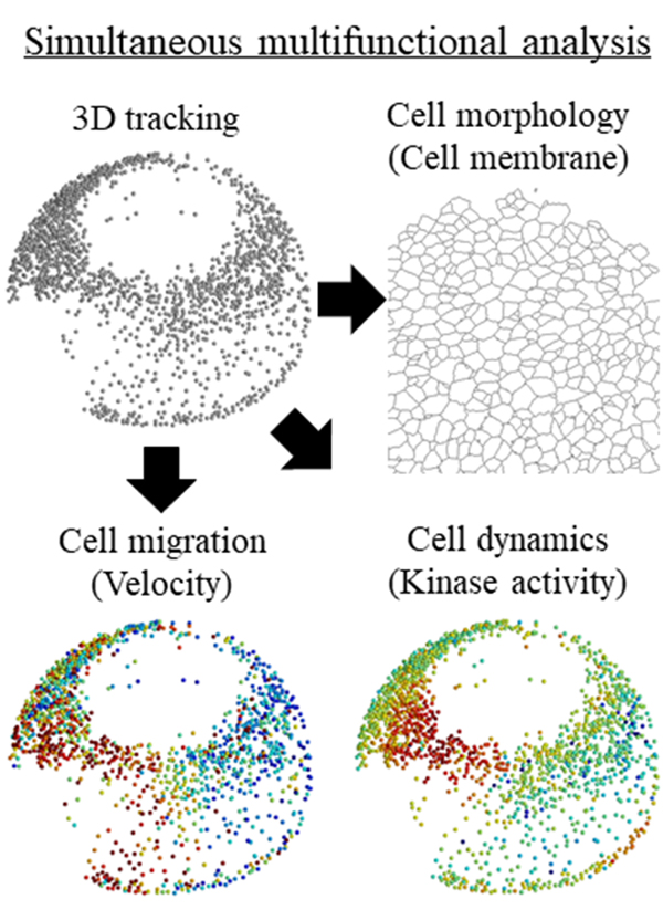
Figure 1. Simultaneous multifunctional analysis of early embryonic development

II. Research support by image analysis

The development of imaging technology in life science research has been remarkable, allowing many researchers to easily acquire large and complex image data sets. However, image analysis is still a hurdle for researchers and can become a research bottleneck. To solve this problem, I provide research support based on the following three concepts.
 
The first concept is quantitative image analysis based on extensive knowledge of image processing and statistics. For most researchers, image evaluation is limited to qualitative and subjective analysis. Correct analysis based on knowledge of image and statistics supports quantitative and objective analysis. The second concept is to support image analysis through machine learning, including deep learning. Machine learning has made remarkable progress in recent years, and with a little learning, it is possible to simplify analysis, which has been difficult with conventional image analysis techniques. The third concept relates to providing researchers with easy-to-understand explanations of image analysis techniques on the Web. The content ranges from the principles of image analysis methods to how to use image analysis software and plug-ins.
 

2021_ohta_en_fig2.jpg
Figure 2. A website that explains how to use image analysis software

Reports

Selected Publications

Yagi, H., Yagi-Utsumi, M., Honda, R., Ohta, Y., Saito, T., Nishio, M., Ninagawa, S., et al. (2020). Improved Secretion of Glycoproteins Using an N-Glycan-Restricted Passport Sequence Tag Recognized by Cargo Receptor. Nat. Commun. 11, 1368.
 
Ohta, Y., Furuta, T., Nagai, T., and Horikawa, K. (2018). Red fluorescent cAMP indicator with increased affinity and expanded dynamic range. Sci. Rep. 8, 1866.
 
Ohta, Y., Kamagata, T., Mukai, A., Takada, S., Nagai, T., and Horikawa, K. (2016). Nontrivial Effect of the Color-Exchange of a Donor/Acceptor Pair in the Engineering of Förster Resonance Energy Transfer (FRET)-Based Indicators. ACS Chem. Biol. 11, 1816-1822.

Contact

E-mail: yohta@nibb.ac.jp