日本語

National Institutes of Natural Sciences

National Institute for Basic Biology

NIBB Departments

Laboratory for Cell Vigor Regulation

Staff

Research Summary

Multicellular organisms build their bodies by repeatedly undergoing cell division starting from a fertilized egg. The cells produced through cleavage change their characteristics depending on their position. The production and removal of signaling molecules that shape body patterns must occur at precisely the right moment within the limited timeframe of development. This timely switching is made possible by the programmed cell death (apoptosis) of the cells of the signal center that produce these signaling molecules.

Additionally, during development, many cells that fail to differentiate properly also appear. Such cells are promptly eliminated from the organism. The mechanisms of cell death are integrated into the creative process of development.

Once development is complete, the body grows rapidly and reaches sexual maturity, enabling reproduction. However, this state does not last indefinitely—organisms cannot live beyond their maximum lifespan. Aging process is also integrated into life history. While organisms adapt to environmental changes or tissue damage to sustain life, a phenomenon known as dormancy, in which development and growth temporarily pause, is also known as a biological response.

By investigating mechanisms that reduce cell or organismal vigor—such as cell death, aging, and dormancy—we aim to deepen our understanding of organismal healthy life history.
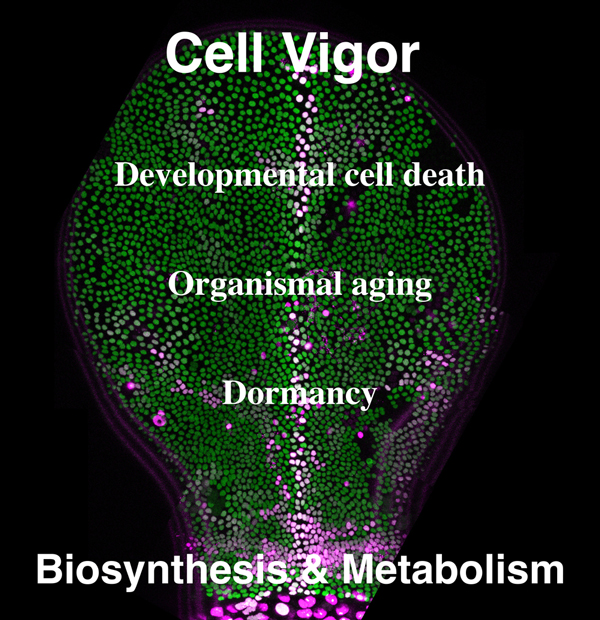
2025_miura_main_visual.jpg

Caspase activation at the midline during thorax closure in Drosophila metamorphosis
During thorax closure in Drosophila metamorphosis, epithelial cells from the left and right sides fuse at the midline, where caspase activation leads to cell extrusion. This cell extrusion plays a role in regulating the speed of closure.

Research Projects

  • • Regulation of cell vigor by methionine/SAM metabolism
  • • Regulation of cell vigor by diphthamide modification of translation elongation factor 2

Selected Publications

Obata, F., and Miura, M. (2024). Regulatory mechanisms of aging through the nutritional and metabolic control of amino acid signaling in model organisms. Annu. Rev. Genet. 58, 19-41.
 
Kosakamoto, H., Obata, F., Kuraishi, J., Aikawa, H., Okada, R., Johnstone, J.N., Onuma, T., Piper, M.D.W., and Miura, M. (2023). Early-adult methionine restriction reduces methionine sulfoxide and extends lifespan in Drosophila. Nat. Commun. 14, 7832.
 
Tsuda-Sakurai, K., and Miura, M. (2019). The hidden nature of protein translational control by diphthamide – the secrets under the leather. J. Biochem. 165, 1-8.
 
Obata, F., Tsuda-Sakurai, K., Yamazaki, T., Nishio, R., Nishimura, K., Kimura, M., Funakoshi, M., and Miura, M. (2018). Nutritional control of stem cell division through S-adenosylmethionine in Drosophila intestine. Dev. Cell 44, 741-751.
 
Kashio, S., Obata, F., Zhang, L., Katsuyama, T., Chihara, T., and Miura, M. (2016). Tissue non-autonomous effects of fat body methionine metabolism on imaginal disc repair in Drosophila. Proc. Natl. Acad. Sci. USA 113, 1835-1840.
 
Obata, F., and Miura, M. (2015). Enhancing S-adenosyl-methionine catabolism extends Drosophila lifespan.  Nat. Commun. 6, 8332.

Obata, F., Kuranaga, E., Tomioka, K., Ming, M., Takeishi, A., Chen, C-H., Soga, T., and Miura, M. (2014). Necrosis-driven systemic immune response alters SAM metabolism through the FOXO-GNMT axis. Cell Rep. 7, 821-833.