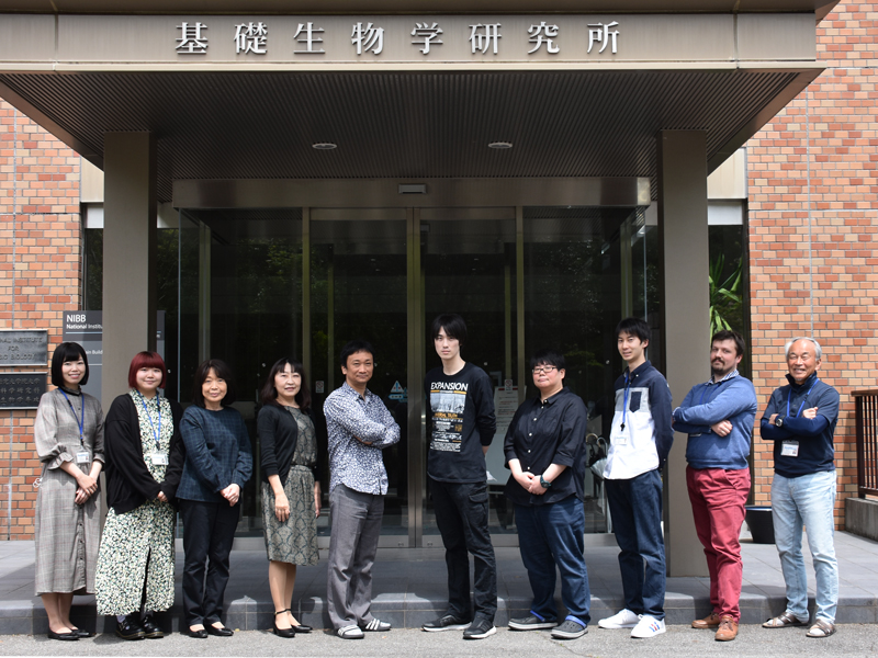
"Why regeneration-competent animals such as newts can restore their missing body parts, but mice and humans can not?” In the past decade, great advances in regeneration studies have revealed many of the molecular mechanisms underlying regeneration. Thus, we now aim to develop strategies to induce regenerative response in humans for replenishing missing tissues and organs by understanding the molecular basis of regeneration from regeneration competent animals.
Research Projects
-
• Elucidating the regulatory mechanisms of adult pluripotent stem cells in planarians.
-
• Inducing frog limb regeneration by substituting certain genomic sequences from frogs with those from newts.
-
• Inducing joint regeneration in mice based on principles of joint regeneration in newts and frogs.
Comparative Regenerative Biology
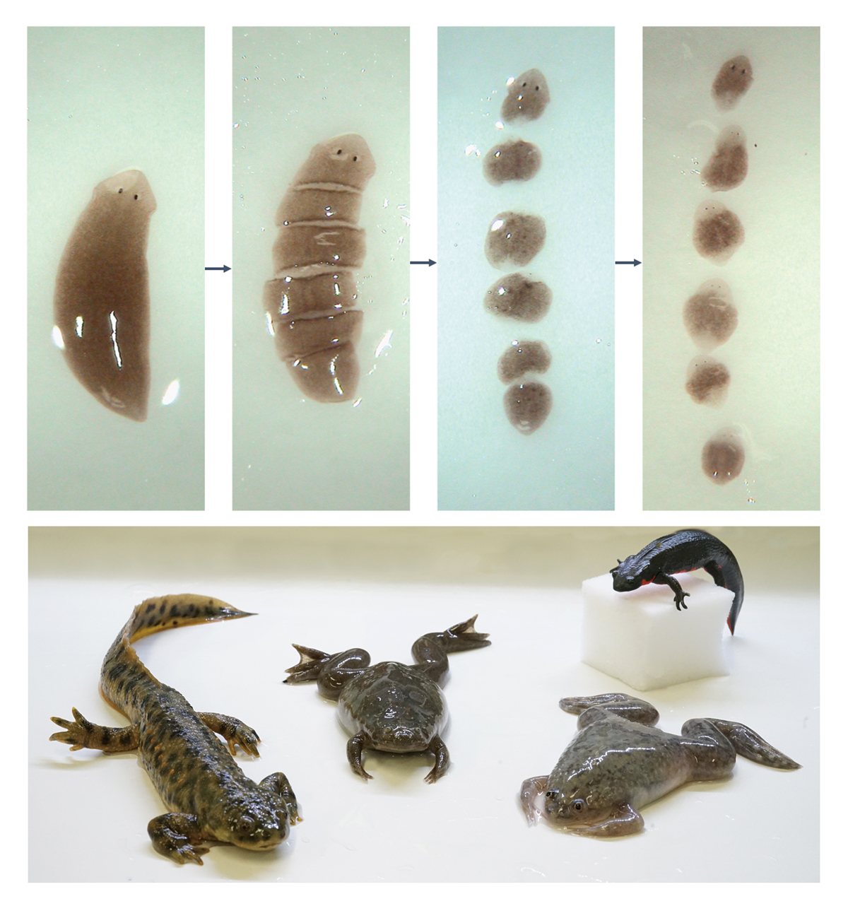
We use animals that demonstrate a high ability in regenerating body parts, such as planarians and newts, to understand the principle of regeneration. In particular, we investigate the difference between regenerative and non-regenerative animals to evoke said abilities from non-regenerative animals. We have already succeeded in achieving this with planarians, which were able to regenerate their heads through RNAi (Umesono
et al., 2013 Nature), and accomplishing functional joint regeneration in frogs through the activation of reintegration systems (Tsutsumi
et al., 2016 Regeneration).
We are currently trying to induce limb regeneration abilities in frogs, as they lose the capability to achieve complete limb regeneration after metamorphosis. We are now focusing on the
Sonic hedgehog (
Shh) super-enhancer MFCS1 (mammals-fishes conserved sequence 1), since it has been suggested that the loss of MFCS1 activity after metamorphosis might cause a failure in achieving the aforementioned limb regeneration in adult frogs (Yakushiji
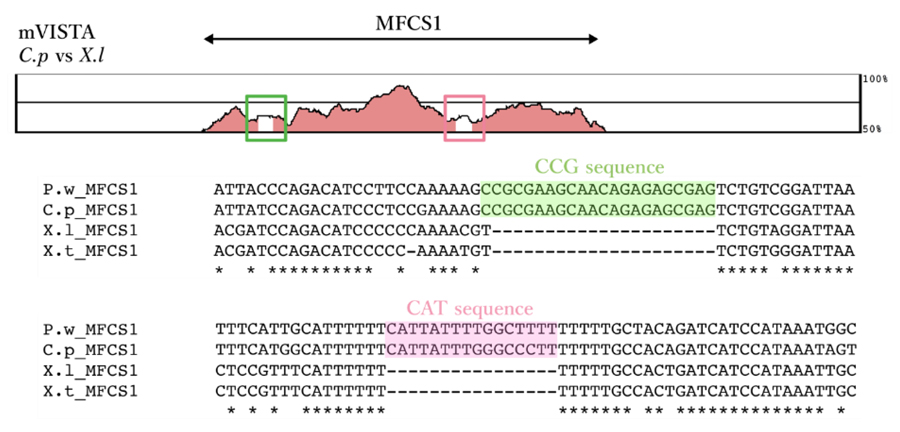
et al., 2009). When we compared the MFCS1 sequences between newts and frogs, newts were found to possess several specific sequences (Figure 1), suggesting that sequence differences might affect super-enhancer formation between newts and frogs after metamorphosis.
Figure.1 Comparison of the MFCS1 sequences between newts and frogs.
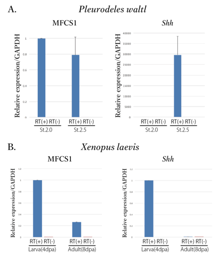
We subsequently tried to detect enhancer RNA (eRNA) which might be transcribed in the MFCS1 region. An interesting aspect of this was that eRNA was detected in regenerating blastema of adult newt from st. 2.0 (
Pleurodeles waltl: Figure 2A).
Conversely, expression of eRNA was suppressed in the frog’s blastema (
Xenopus laevis) after metamorphosis (Figure 2B).
Figure.2 Expression of MFCS1-eRNA and
Shh mRNA in the regenerating blastema of newt (
P.w.) and frog (
X.l.)
(A) The left and right panels show expression levels of MFCS1-eRNA and
Shh mRNA, respectively. The left and right graphs in each panel were obtained from regenerating blastema at St.2.0 and St.2.5 of adult newts after amputation.
(B) The left and right panels shows expression levels of MFCS1-eRNA and
Shh mRNA in frogs before and after metamorphosis.
Trials in isolating viable adult pluripotent stem cells derived from planarian using FACS
We have tried to develop an isolation method for viable adult pluripotent stem cells (aPSC) from planarians using FACS and succeeded in conditioning the low toxic staining method with both nuclear and cytoplasimic fluorescence dyes. 1µM Hoechst 33342 or 0.05 µg/ml Calsein AM could be used for isolating viable aPSC of the planarian,
Dugesis japonica. We are presently investigating the effects of FGF- and Wnt-morphogens to cultured aPSC to demonstrate “the double gradient hypothesis”, which was proposed by Thomas Hunt Morgan (Morgan, 1901) and our group (Umesono
et al., Nature, 2013).