|
|
 |
Title: |
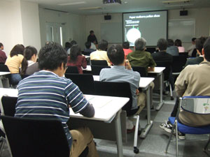
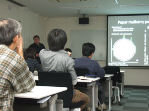
Light sheet based Fluorescence Microscopes (LSFM, SPIM, DSLM) - Tools for a modern biology |
| |
Speaker: |
Dr. Ernst Stelzer
Group Leader, Cell Biology and Biophysics Unit, EMBL Heidelberg, Germany
|
| |
Place: |
Myodaiji Conf. Rm. (111) |
| |
Date: |
2008.04.17(Thu) 16:00 - 17:30 |
| |
Abstract: |
The resolution of optical microscopes is in the range of several 100 nm, the precision of optical tweezers can be as low as a single nm [1], laser cutters such as the laser nanoscalpel generate incisions a couple of 100 nm wide and in three dimensions cause severing that is barely 700 nm deep [2]. More recently, extremely efficient light microscopes require only nanoWatts of power per volume element to induce a reasonably detectable fluorescence emission [3].
Surprisingly, most optical technologies are applied to 2D cellular systems, i.e. in a cellular context that is defined by hard and flat surfaces. However, meaningful information relies on the morphology, the mechanical properties, the media flux and the biochemistry of a cell's context as found in live tissues [4, 5]. Such a physiological context is not found in single cells cultivated on cover slips. It requires a more complex three- dimensional construct of cells, patterned surfaces, and cultivation in an ECM-based gel or in embryos of flies and fish or live tissue [5].
The observation as well as the optical manipulation of thick and optically dense biological specimens suffers from at least two severe problems. 1) The specimens tend to scatter and absorb light. The delivery of the probing light as well as the collection of the signal light tends to become inefficient. 2) Many biochemical compounds apart from the fluoro-phores also absorb light and suffer degradation of some sort (photo toxicity), which induces malfunction or death of a specimen [3].
We develop technologies for the observation and manipulation of large and complex three-dimensional biological specimens. The basic technol- ogy of choice seems to be the use of light sheets, which are fed into the specimen from the side and observed at an angle of 90° to the illumination optical axis. The focal volumes of the detection lens and the volume of the light sheet overlap. Thus, optical sectioning and no photo damage outside the common focal plane become intrinsic properties.
EMBL's novel microscopes (SPIM, DSLM) take advantage of modern camera technologies and can be combined with essentially every contrast [6] and specimen manipulation tool. However, they operate in a truly three-dimensional fashion. The straightforward optical path in LSFM is designed to allow high flexibility and modularity. In a recent application, DSLM is used to record fish embryonic development in vivo and in toto, from very early stages until late neurulation with subcellular resolution and short sampling periods. We follow the cell movements during gas-trulation and reveal important development during the migration processes. The perspective is to continue to use our technology in embryology as well as in tissue engineering, e.g. in the development of physiologically relevant toxicology assays. We are also convinced that our truly three-dimensional approach will
have a dramatic impact on biophysical studies [7].
References
[1] Trapping and tracking a local probe with a PFM.Rev. Sci. Instr. 2004 75:2197-2210.
[2] Investigating relaxation processes in cells and developing organisms.Methods in Cell Biology (Edt. Berns M & Greulich KO), 2007 82:267-91.
[3] Optical sectioning deep inside live embryos by SPIM. Science 2004 305:1007-9.
[4] 3D prepara-tion and imaging reveal intrinsic MT properties, Nat Methods, 2007 4(10):843-846.
[5] The third dimension bridges the gap between cell culture and live tissue. Nat Rev MCB 2007 8(10):839-845.
[6] High-resolution 3D imaging of large specimens with light-sheet based microscopy. Nat Methods, 2007 4:311-313.
[7] Three-dimensional microtubule behaviour in Xenopus egg extracts reveals four dynamic states and state-dependent elastic properties. Biophys. J. 2008 in press.
|
|


 Top Page
  Symposium/Meeting
  EMBL Guest Seminar
  Research Collaboration
  Students Communication


