NATIONAL INSITUTE FOR BASIC BIOLOGY  


National Institute for Basic Biology

DIVISION OF CELL DIFFERENTIATION


Professor:
Ken-ichirou Morohashi
Research Associate:
Satoru Ishihara
Technical Staff:
Sanae Oka
Post Doctral Fellow:
Naoe Kotomura
Graduate Students:
Ken Kawabe (Kyushu University)
Tokuo Mukai (Kyushu University)
Hirofumi Mizusaki (The Graduate University for Advanced Studies)
Tatsuji Shikayama (Kyushu University)
Hayato Yokoi (Nagoya University)
Akira Nanba (University of Tokyo)
Ryoji Matsushima (Nagoya University)
Visiting Fellow:
Yoshiyuki Kojima (Nagoya City University)
Tetsushi Dodo (Eisai Co. Ltd)
Assistant:
Hisae Tsuboi


Steroid hormones, which act as endocrine messengers, are mainly synthesized in steroidogenic tissues such as the gonads and adrenal cortex. Because of the pivotal function of sex steroids and corticosteroids secreted from these tissues, several groups have investigated the regulation of hormone production. Specifically, in the past decade, extensive research has been directed into the area of transcriptional regulation of genes implicated in steroidogenesis. As one of these studies, we identified Ad4BP/SF-1, which belongs to a nuclear hormone receptor family, as a major steroidogenic tissue-specific transcription factor. Further studies have suggested that this versatile transcription factor is central to the regulation of a reproductive behavior thorugh functioning in the pituitary gonadotroph and ventromedial hypothalamic nucleus in addition to the steroidogenic tissues. Based on these observation, our attention has been directed to understanding mechanisms underlying differentiation of the reproductive system and establishment of reproductive behavior of animals.


I. Transcriptinal control of genes necessary for gonadal differentiation.

Because of the fundamental and pivotal function of the gonads, extensive efforts have been made to characterize the differentiation processes including sex-dependent differentiation. By focusing on the steroidogenesis as one of the gonad specific functions, we identified a steroidogenic tissue specific transcription factor designated Ad4BP/SF-1. Recent studies with an antiserum to Ad4BP/SF-1 and a gene disrupted mice clearly demonstrated that the transcription factor is essential for differentiation of the steroidogenic tissues, the gonads and the adrenal cortex. Showing a good correlation with the observation, a particular cell population immunoreactive for Ad4BP/SF-1 (AGP, adreno-gonadal primordium) was identified to be present from the dorsal aorta to coelomic epithelia of rodent fetauses and succesively they were shown to give rise to two distinct primordia, the gonads and adrenal cortex. In the case of the gonadal primodium, it finally gives rise to the testis or ovary according to the sex chromosomal composition. These observations gave us many interesting issues as follows. What is the signal for the onset of the Ad4BP/SF-1 gene transcription in the particular cells comprising AGP? What is the mechanism underlying the separation of the AGP into the adrenal and gonadal primordia? How is the mechanism employed for sex differentiation of the gonads? Why dose such sex dependent differentiation not occur in the adrenal cortex? To address these issues, nuclear transcription factors (WT-1, SRY, SOX-9, DAX-1, Emx-2, and GATA-4, as well as Ad4BP/SF-1), all of which are critically implicated in gonadal and adrenocortical differentiation, havebeen underinvestigation from the molecular and morphological aspects.

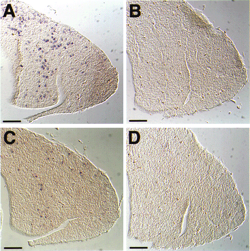
As one of these efforts, we investigsted Dax-1 gene regulation and identified a novel Ad4 site, which is recognized by Ad4BP/SF-1, by transient transfection and electrophoretic mobility shift assays. In addition, immunohistochemical analyses with antibodies specific for Dax-1 indicated the presence of immunoreactive cells in the steroidogenic tissues, pituitary gland and hypothalamus. Although the distributions of Dax-1 and Ad4BP/SF-1 were highly similar, they were not completely identical. Namely a certain population of cells immunoreactive for Ad4BP/SF-1 is negative for Dax-1 expression. Such inconsistent distributions between the two transcription factors were observed in all tisseus. To confirm the positive regulation of the Dax-1 gene by Ad4BP/SF-1, the Dax-1 expression was investigated with the mFtz-F1 gene disrupted mice. The Dax-1 expression was significantly impaired in the mFtz-F1 gene disrupted mice (Fig. 1), confirming our understanding that Ad4BP/SF-1 controls the transcription of the Dax-1 gene.

Fig.1
Immunohistochemical detection of Dax-1 in the pituitary. Sections of the pituitary glands from the newborn were prepared from wild-type (A and C) and mFtz-F1 gene disrupted (B and D) mice. They were stained with anti-Ad4BP/SF-1 antisera (A and B) or anti-Dax-1 antibodies (C and D) as described. Bars; 50 mm.


II. Implication of Ad4BP/SF-1 in strctural organization of spleen.

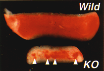
D uring characterization of the mFtz-F1 gene disrupted mice, we were aware of the abnormal spleen of the mFtz-F1 gene disrupted mice in addition to the tissues above (Fig. 2). The spleen has normally two main functions. The first is to provide a proper microenvironment to lymphoid and myeloid cells, while the second involves clearance of abnormal, damaged, and aged erythrocytes. Immuno-histochemical examination of the mammalian spleens confirmed the expression of Ad4BP/SF-1 in endothelial cells of the splenic venous sinuses and pulp vein. In the mFtz-F1 gene disrupted mice, several structural abnormalities were detected in the spleen, including underdevelopment and nonuniform distribution of erythrocytes. Examination of the spleen of KO fetuses showed failure of development of certain tubular structures during embryogenesis. These structures are normally assembled by Ad4BP/SF-1 immunoreactive cells, and most likely form the vascular system during later stages of development. Other structural abnormalities in the spleen of the mFtz-F1 gene disrupted mice included defects in the tissue distribution of type IV collagen, laminin, c-Kit, and vimentin. These morphological defects in the vascular system were associated with a decrease in the proportion of hematopoietic cells although differentiation of these cells was not affected significantly. A high number of abnormal red blood cells containing Howell-Jolly bodies were noted in the mFtz-F1 gene disrupted mice, indicating impaired ability for clearance due to the affected splenic vascular system. We also detected the presence of an mRNA encoding cholesterol side chain cleavage P450 in the spleen, resembling the findings in steroidogenic tissues such as the gonads and adrenal cortex. The mRNA transcript was not involved in splenic structural defects as it was detected in the spleens of both normal and the mFtz-F1 gene disrupted mice, indicating that the regulatory mechanism of the P450 gene in the spleen is different from that in steroidogenic tissues. Our results indicate a lack of the mFtz-F1 gene in mice is associated with structural and functional abnormalities of the splenic vascular system (In press, Blood, 1999).

Fig.2
Comparison of macroscopic features of spleens of wild-type (Wild) and mFtz-F1 gene disrupted (KO) newborn mice. Arrowheads show red spots characteristic of the KO spleen.


Publication List:
Kawano, K., Miura, I., Morohashi, K., Takase, M., and Nakamura, M. (1998) Molecular cloning and expression of the SF-1/Ad4BP gene in the frog, Rana rugosa. Gene 222, 169-176.
Nomura, M., Kawabe, K., Matsushita, S., Oka, S., Hatano, O., Harada, N., and Nawata, H., and Morohashi, K. (1998) Adrenalcortical and gonadal expression of the mammalian Ftz-F1 gene encoding Ad4BP/SF-1 is independent of pituitary control. J. Biochem. 124, 217-224.


webmaster@nibb.ac.jp
Last Modified: 12:00, May 28, 1999