NATIONAL INSITUTE FOR BASIC BIOLOGY  


National Institute for Basic Biology

DIVISION OF REPRODUCTIVE BIOLOGY


Professor:
Yoshitaka Nagahama
Associate Professor:
Michiyasu Yoshikuni
Research Associates:
Minoru Tanaka
Tohru Kobayashi
Institute Research Fellow:
Akihiko Yamaguchi
JSPS Postdoctoral Fellows:
Mika Tokumoto
Yoshinao Katsu
Daisuke Kobayashi
Craig Morrey
Catherine Dreanno
Balasubramarian Senthilkumaran
JSPS Research Associates:
Yuichi Ohba
Yasutoshi Yoshiura
Masaru Matsuda
Toshitaka Ikeuchi
Graduate Students:
Masatada Watanabe (Graduate University for Advanced Studies)
Gui-Jin Guan (Graduate University for Advanced Studies)
Ryo Horiuchi (Graduate University for Advanced Studies)
Monbusho Foreign Scientist:
Allen Schuetz (Johns Hopkins University)
Srinivas K. Saidapur (Karnatak University)
Visiting Scientist: Graham Young (University of Otago)


Gametogenesis of teleost fishes, as in other oviparous vertebrates, is controlled primarily by pituitary gonadotropins. In most cases, gonadotropins act via biosynthesis of gonadal steroidal hormones which, in turn, mediate various stages of gametogenesis. Using fish as a primary study model, our research focuses on (1) the identification of steroidal mediators involved in gonadal sex differentiation and gametogenesis, and (2) the mechanisms of synthesis and action of these mediators.


I. Endocrine regulation of oocyte growth and maturation

Two follicular steroidal mediators, estradiol-17b (oocyte growth) and 17a,20b-dihydroxy-4-pregnen-3-one (17a,20b-DP; oocyte maturation), were identified in several teleost fishes. Two-cell type models in which the thecal layer provides precursor steroids to the granulosa layer, have been proposed for estradiol-17b and 17a,20b-DP production. There is a distinct shift in expression of steroidogenic enzyme genes from cytochrome P450 aromatase (P450arom) to 20b-hydroxysteroid dehydrogenase (20b-HSD) in granulosa cells immediately prior to oocyte maturation. The presence of orphan nuclear binding sites in the medaka P450arom promoter and the similar expression patterns of FTZ-F1 and P450arom transcripts in ovarian follicles suggest that FTZ-F1 is involved in the transcriptional regulation of medaka P450arom during oocyte growth. The preovulatory surge of LH-like gonadotropin is responsible for the rapid expression of 20b-HSD mRNA transcripts in granulosa cells during oocyte maturation.

17a,20b-DP induces oocyte maturation by acting on a pertussis toxin-sensitive G-protein-coupled membrane receptor. The early steps of l7a,20b-DP action involve the formation of downstream mediator of this steroid, the maturation-promoting factor or metaphase-promoting factor (MPF) consisting of cdc2 kinase and cyclin B. 17a,20b-DP induces oocytes to synthesize cyclin B which activates a preexisting 35-kDa cdc2 kinase via phosphorylation of its threonine 161 by a threonine kinase (MO15), thus producing the 34 kDa active cdc2. A 54-kDa Y box protein and polyadenylation of cyclin B mRNA are thought to be involed in 17a,20b-DP-induced initiation of cyclin B mRNA translation.

U pon egg activation, MPF is inactivated by degradation of cyclin B. The availability of biologically-active goldfish cyclin B produced in E. coli and purified goldfish proteasomes allowed the role of the proteasome in the regulation of cyclin degradation to be examined for the first time. It was demonstrated that the 26S proteasome initiates cyclin B degradation through the first cut of its NH2 terminus at lysine 57.


II. Endocrine regulation of spermatogenesis

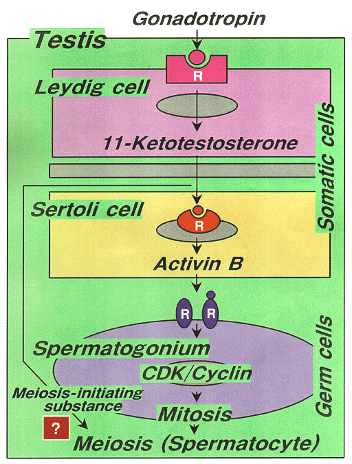
The importance of gonadotropins and androgens for spermatogenesis is generally accepted in vertebrates; however, the mechanism of action of these hormones remains unresolved. Using an organ culture system for eel testes consisting of spermatogonia and inactive somatic cells, we have shown that the hormonal regulation of spermatogenesis in eel testes involves the gonadotropin stimulation of Leydig cells to produce 11-ketotestosterone (11-KT), a potent androgen in fish. In turn, 11-KT activates Sertoli cells to stimulate premitotic spermatogonia to complete spermatogenesis (Fig. 1). A subtractive hybridization method was used to clone genes that are expressed differentially in eel testes in the first 24 hr after gonadotropin treatment. Six down-regulated and two up-regulated cDNA clones were isolated. Two cDNA clones suppressed by gonadotropin stimulation exhibited ZP (zona pellucida sperm-binding protein)2- and ZP3-like structures. Transcripts of ZP2- and ZP3 are detected in immature testes and disappear immediately after HCG injection, suggesting that ZP2 and ZP3 may play important roles in the prevention of spermatogenesis in the eel. One of the up-regulated cDNAs was identified as coding the activin bB subunit. Activn bB mRNA transcripts were absent in testes prior to gonadotropin treatment, but were abundant in Sertoli cells in testes of eels treated with gonadotropin for 1-6 days. 11-KT induced a marked production of activin B in cultured testes, indicating that eel activin bB subunit production is largely for activin B formation. An androgen receptor localized in Sertoli cells. Addition of recombinant eel activin B to the culture medium induced proliferation of spermatogonia, producing late type B spermatogonia, within 15 days in the same manner as did 11-KT. Activin B also acts on spermatogonia to induce de novo synsthesis of G1/S cyclins (cyclins A, D, and E) and Cdks (cdc2, cdk2, and cdk4), leading to the initiation of mitosis (spermatogonial proliferation) (Fig. 1).

Fig. 1
Hormonal regulation of spermatogenesis initiation in the Japanese eel.


III. Endocrine regulation of gonadal sex differentiation and sex change

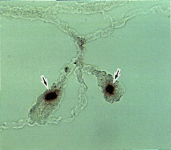
The initial differentiation and development of steroid-producing cells during gonadal sexual differentiation were examined by in situ hybridization and immunohistochemistry in tilapia, Oreochromis niloticus. Transcripts of P450arom became evident for the first time in the gonads of genetic females two weeks before morphological sex differentiation (Fig. 2). These gonads were also positive to antibodies against P450scc, 3b-HSD, P450c17, and P450arom. Thus, steroid-producing cells in ovaries, but not testes, at the undifferentiated and differentiating stages express all of the steroidogenic enzymes required for estradiol-17b biosynthesis from cholesterol. These results strongly suggest that endogenous estrogens act as the natural inducers of ovarian differentiation in tilapia. In contrast, weakly positive reactions to P450scc, 3b-HSD and P450c17 antibodies appeared first in the testis at 30 days; however, staining intensity increased slowly during development of the testis. Therefore, the ability of steroid-producing cells to synthesize steroid hormones in the testes only appears at the time of testicular differentiation.

Fig. 2
Expression of cytochrome P450 aromatase mRNA in the gonad of tilapia two weeks before morphological sex differentiation. Note strong signals in steroid-producing cells (arrows).

Sequential, protogynous hermaphroditism, i.e. female to male sex change, provides an ideal comparative model for studies on sexual differentiation. Fully-functional females are induced to develop functional testes by non-invasive social conditions, thus allowing simultaneous investigation into the normal endocrine requirements of ovarian maintenance and testicular differentiation. As steroid hormones are considered major mediators of sexual differentiation and gonadal function, molecular studies of key enzymes in the steroidogenic pathways were completed. Two different P450arom mRNAs as well as P450 11b-hydroxylase mRNA have been cloned and characterized throughout sex change in Thalassoma duperrey, a protogynous hermaphrodite. The ovarian form of P450arom is predominantly expressed in the ovary prior to the initiation of sex change. Shortly thereafter, expression diminished to undetectable levels suggesting that ovarian P450arom/estrogen is critical for the maintenance of ovarian function and/or detrimental to testicular differentiation. Although intriguingly complex and potentially relevant, the neural form of P450arom, named for its abundance in the brain, does not appear to be directly related to the gonadal restructuring seen during sex change. Nevertheless, its function and significance are being actively pursued. In contrast, P450 11b-hydroxylase is likely to be a major factor regulating the gonadal changes. P450 11b-hydroxylase mRNA was relatively abundant in the ovary; however, it was up-regulated concomitant with the onset of testicular differentiation indicating the importance of P450 11b-hydroxylase/11-KT to spermatogenesis and spermiation. Consequently, the switch from an ovary to a testis corresponds exceptionally well to a switch in the steroidogenic pathway of the gonad thus providing further evidence of the critical nature of steroid hormones in teleost sexual differentiation.


Publication List:

Horiguchi, R., Tokumoto, M., Yoshiura, Y., Aida, K., Nagahama, Y. and Tokumoto, T. (1998). Molecular cloning of cDNA encoding a 20S proteasome a2 subunit from goldfish (Carassius auratus) and its expression analysis. Zool. Sci., 15, 773-777.
Jiang, J.Q., Young, G., Kobayashi, T. and Nagahama, Y. (1998). Eel (Anguilla japonica) testis 11b-hydroxylase gene is expressed in interrenal tissue and its product lacks aldosterone synthesizing activity. Mol. Cell. Endocrinol., 146, 207-211.
Kobayashi, T., Kajiura-Kobayashi, H. and Nagahama, Y. (1998). A novel stage-specific antigen is expressed only in early stages of spermatogenesis in Japanese eel, Anguilla japonica testis. Mol. Reprod. Develop., 51, 355-361
Kobayashi, T., Nakamura, M., Kajiura-Kobayashi, H., Young, G. and Nagahama, Y. (1998). Immunolocalization of steroidogenic enzymes (P450scc, P450c17, P450arom, and 3b-HSD) in immature and mature testes of rainbow trout (Oncorhynchus mykiss). Cell Tissue Res., 292, 573-577.
Mita, M., Yasumasu, I., Saneyoshi, M., Yoshikuni, M. and Nagahama, Y. (1998). Production of the oocyte maturation-inducing substance of starfish by heat treatment of S-adenosylmethionine. Zool. Sci., 15, 117-122.
Mita, M., Yoshikuni, M. and Nagahama, Y. (1998). Ecto-ATP diphosphohydrolase (apyrase) in ovarian follicle cells of starfish Asterina pectinifera. Comp. Biochem. Physiol. Part B, 119, 577-583.
Miura, T., Kudo, N., Miura, C., Yamauchi, K. and Nagahama, Y. (1998). Two testicular cDNA clones suppressed by gonadotropin stimulation exhibit ZP2- and ZP3-like structures in Japanese eel. Mol. Reprod. Develop., 51, 235-242.
Morrey, C.E., Nakamura, M., Kobayashi, T., Grau, E.G. and Nagahama, Y. (1998). P450scc-like immunocytochemistry throughout gonadal restructuring in the protogynous hermaphrodite Thalassoma duperrey. Int. J. Develop. Biol., 42, 811-816.
Nakamura, M, Kobayashi, T., Chang, X.T. and Nagahama, Y. (1998). Gonadal sex differentiation in teleost fish. J. Exp. Zool., 362-372.
Parhar, I.S., Nagahama, Y., Grau, E.G. and Ross, R.M. (1998). Immunocytochemical and ultrastructural identification of pituitary cell types in the protogynous Thalassoma dupperrey during adult sexual ontogeny. Zool. Sci., 15, 263-276.
Parhar, I.S., Soga, T., Ishikawa, Y., Nagahama, Y. and Sakuma, Y. (1998). Neurons synthesizing gonadotropin-releasing hormone mRNA subtypes have multiple developmental origins in the medaka. J. Comp. Neurol., 401, 217-226.
Todo, T., Ikeuchi, T., Kobayashi, T. and Nagahama, Y. (1998). Fish androgen receptor: cDNA cloning, steroid activation of transcription in transfected mammalian cells, and tissue mRNA levels. Biochem. Biophys. Res. Comm., 254, 378-383.


webmaster@nibb.ac.jp
Last Modified: 12:00, May 28, 1999