NATIONAL INSITUTE FOR BASIC BIOLOGY  


National Institute for Basic Biology

Division of Morphogenesis


Professor:
Goro Eguchi
Associate Professor:
Ryuji Kodama
Research Associates:
Makoto Mochii
Mitsuko Kosaka
Visiting Scientists:
Takamasa S. Yamamoto
Akio Iio (from Chubu National Hospital)
Shin-ichi Higashijima (from Japan Science and Technology Corporation)
Panagiotis A. Tsonis (from University of Dayton)
Katia Del Rio-Tsonis (from University of Dayton)
Thomas Reh (from University of Washington)
Graduate Student:
Harutoshi Hayashi (from School of Agriculture, University of Tokyo)
Technical Staffs:
Chiyo Takagi
Sanae Oka


In the newt and some other limited animal species, the lens and neural retina can be completely regenerated through transdifferentiation of pigmented epithelial cells (PECs). Such a phenomenon, transdifferentiation, as observed in regeneration of ocular tissues seems to be a highly powerful model for studying stability and instability in differentiation of tissue cells. From this view point, lens transdifferentiation of PECs of vertebrate has been studied using in vivo and in vitro systems, and our in vitro studies have revealed that dormant potential of PECs to transdifferentiate into lens cells is widely conserved throughout vertebrate species and that such potential becomes evident when PECs are cultured in vitro. Such transdifferentiation can be accelerated in the presence of phenylthiourea and testicular hyaluronidase and cells passes through a dedifferentiated status, where expression of PEC-specific genes, such as mmp115, tyrosinase, TRP-1, and pP344 are suppressed. On the other hand, c-myc and pax6 expressions are elevated in the dedifferentiated PECs. We have extended our study to the regulatory mechanisms controlling gene expression during transdifferentiation and following results are obtained.


I. In vitro culture system of iris pigmented epithelial cells for molecular analysis of lens regeneration

We have succeeded in purely isolating and cultivating the iris pigmented epithelial cells of 2 day-old chick using chemical treatment by dispase and EDTA. The iris PE cells continued to proliferate very stably in the control medium and transdifferentiate into lens cells by the addition of only two defined growth factors to the culture medium. The process of transdifferentiation in this system is very reproducible and this culture system of the iris PE has made it possible to prepare homogeneous cell population at various stages during the transdifferentiation process, thus providing the way for biochemical and molecular biological analyses. The Northern blot analysis suggests that the transcription level of pax6 gene closely correlate with the progression of the transdifferentiation process of the iris PE cells to the lens.


II. Role of the mi gene in differentiation and transdifferentiation of the pigmented epithelial cells

The mi gene was first isolated as a mouse gene in the microphthalmia (mi) locus and shown to encode a basic-helix-loop-helix-leucine zipper (bHLHzip) transcription factor, which is a possible regulatory gene functioning in PEC differentiation. We have revealed that mi is specifically expressed in PECs and precursors of PECs in the embryo, and that mi starts to be expressed at the optic vesicle stage when no other marker genes are expressed yet. Expression of mi is also correlated with PEC differentiation in the culture condition, where mi expression is down-regulated under the condition for dedifferentiation but up-regulated under the condition for redifferentiation.

In order to elucidate the role of mi in PEC differentiation, cultured PECs were infected with a retrovirus construction including mi sequence. The mi overexpression did not cause significant changes in morphology but caused a dramatic change in the pattern of gene expression. PECs infected with the retrovirus encoding mi continued to express mmp115, tyrosinase and TRP-1 but not pP344 even under a culture condition which promote dedifferentiation. Transfection and CAT assay revealed that mi transactivates mmp115 promoter. These results suggest that mi has a critical role in PEC differentiation by activating mmp115, tyrosinase and TRP-1. Moreover, the result that pP344 was not transactivated by mi expression suggested the presence of another regulatory factor in PEC differentiation which is required to promote pP344 expression. Our findings revealed that at least a part of the changes in gene expression pattern accompanying dedifferentiation is regulated by mi inactivation which is induced by dedifferentiation signals.


III. Analysis of silver quail mutant

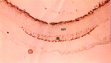
In the homozygous silver mutant of Japanese quail (Coturnix coturnix japonica), a part of the outer layer of retina, which normally differentiates into the pigmented epithelium of the retina, forms an ectopic neural retina tissue (Fig. 1). The ectopic neural retina was suggested to be formed through transdifferentiation from pigmented epithelium according to morphological observations, but there have been no direct evidence. We found that the outer layer of retina in silver homozygote once differentiates to pigmented epithelium judging from the expression of mmp115 in 4-day embryo. After 5th day of incubation, however, the outer layer starts to express neural marker genes which indicates the beginning of the transdifferentiation into the neural retina. The process to form the ectopic neural retina in silver embryo is, therefore, a transdifferentiation process. Furthermore, we analysed the sequence of mi gene in the silver homozygote and found that there is a two-base deletion which causes a production of truncated mi protein. We have confirmed using an in vitro assay that the truncated mi protein has less activity in activating the mmp115 promoter. These results suggest that the mutation in the mi gene induces transdifferentiation of PECs to neural retina cells in silver homozygote.

Fig. 1.
Ectopic neural retina in silver mutant quail.
Retinal section of 7-day-old embryo from silver homozygote was stained with anti-neurofilament antibody. Ectopic neural retina formed via transdifferentiation of pigmented epithelium is positively stained (*).
NR: original neural retina


IV. Isolation of a novel chick homologue of Serrate and its coexpression with C-Notch-1 in chick development

Intercellular signaling mediated by the transmembrane proteins, Notch as receptor and its ligands, Delta and Serrate, plays essential roles in the developmental fate decision of many cell types in Drosophila. The Notch genes are highly conserved both in invertebrates and in vertebrates, suggesting that Notch pathway regulates cell fate decisions during vertebrates development.

As a starting point of our study on the role of the Notch pathway in the control of the differentiation of ocular tissues, we cloned homologues of Notch, Delta and Serrate in the chicken. Among them, there was a novel chick homologue of Drosophila Serrate, named as C-Serrate-2. We examined the expression patterns of C-Serrate-2 and other homologs during the early chick development using whole-mount in situ hybridization. Tissues with conspicuous expression of Notch receptors and ligands included the forebrain, the myotome and the apical ectodermal ridge (AER) of the limb bud of a 4-day-old chick embryo.

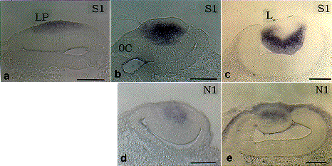
In the early development of ocular tissues, C-Serrate-1 expression was observed in the thickened ectoderm of the lens primordium at stage 13 (Fig. 2). At stage 14, C-Notch-1 transcripts were coexpressed with C-Serrate-1 in the lens placode. When the lens placode invaginates to form a lens vesicle in 3-day-embryos (stage 18-21), C-Serrate-1 expression was restricted to the posterior region of the lens containing differentiating lens fiber cells. On the other hand, C-Notch-1 was restricted to the anterior region of the lens containing proliferating cells.

These combination of expression patterns during developmental processes is useful in predicting possible roles and mode of functions of intercellular signalling through the Notch pathway.

Fig. 2.
Expression of C-Serrate-1 and C-Notch-1 in the eye primordium of the chick embryo.
Embryos at stage 13 (a), stage 14 (b, d) and stage 18-21 (c, e) were hybridized with C-Serrate-1 (S1) or C-Notch-1 (N1) probes.
LP: lens placode, L: lens, OC: optic cup, the scale bar: 100 mm


Publication List:
Mazaki, Y., Mochii, M., Kodama, R. and Eguchi, G. (1996) Role of integrins in differentiation of chick retinal pigmented epithelial cells in vitro. Develop. Growth Differ. 38, 429-437.
Ono, T., Murakami, T., Tanabe, Y., Mizutani, M., Mochii, M. and Eguchi, G. (1996) Culture of naked quail (Coturnix coturnix japonica) ova in vitro for avian transgenesis: culture from the single-cell stage to hatching with pH-adjusted chicken thick albumen. Comp. Biochem. Physiol. 113A(3), 287-292.
Sawada, K., Agata, K. and Eguchi, G. (1996) Characterization of terminally differentiated cell state by categorizing cDNA clones derived from chicken lens fibers. Int. J. Dev. Biol. 40, 531-535.
Furukawa, K., Yamamoto, T., Takamune, K., Sugimoto, Y., Eguchi, G. and Abe, S.-I. (1996) Genesis of newt sperm axial fiber: cDNA cloning and expression of a 29 kDa protein, a major component of the axial fiber, during spermatogenesis. Int. J. Dev. Biol. 40(6), 1109-1118.
Hayashi, H., Mochii, M., Kodama, R., Hamada, Y., Mizuno, N., Eguchi, G. and Tachi, C. (1996) Isolation of a novel chick homolog of Serrate and its coexpression with C-Notch-1 in chick development. Int. J. Dev. Biol. 40(6), 1089-1096.


nibb-adm@nibb.ac.jp
Last Modified: 12:00, June 27, 1997