OFFICE OF DIRECTOR
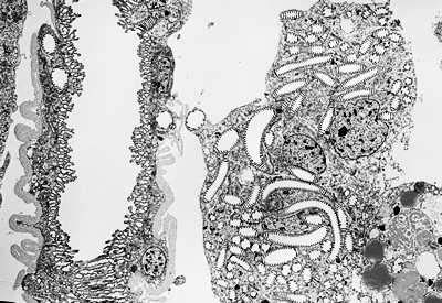
Mechanisms working in the morphogenesis of the lepidopteran wingsKODAMA, Ryuji Wings of the lepidopteran insects (butterflies and moths) develop from the wing imaginal disc, which is a hollow sac made of simple epithelium. Due to its simple construction, this tissue is a good material to study cellular interactions in the course of morphogenesis. The outline shape of the adult wing is often different from that of the pupal wing. This difference is brought about by the programmed cell death of the marginal area of the pupal wing. The marginal dying area is called the degeneration region and the internal area, which develop into the adult wing, is called the differentiation region. The cell deaths in the degeneration region proceeds very rapidly and completes in a half to one day period in Pieris rapae or several other species examined. It was shown that the dying cells in the degeneration region have two characteristics common with the apoptotic cell death in mammalian cells. These are i) the presence of apoptotic bodies, which are heavily condensed cells or their fragments engulfed by other cells or macrophages, shown by transmission electron microscopy and ii) the presence of conspicuous accumulation of fragmented DNA evidenced by the TUNEL histological staining. The cells in the degeneration region are actively engulfed by the macrophages in the cavity beneath the wing epithelium. The macrophages seem to be concentrated beneath the degeneration region by the strong adhesion between basal surfaces of the dorsal and ventral epithelium in the differentiation region. A collaborative work with the laboratory of Dr. K. Watanabe (Hiroshima University) concerns mostly on the development of trachea and tracheole pattern in the swallow tail butterflies. Trachea and trcheoles are both important in delivering air into the wing and their pattern coincide with that of the boundary of degeneration and differentiation zones at the distal end of the wing. According to the observations, the pattern formation of wing epithelium is often dependent on tracheal and tracheole patterns. Basic research on the development of tracheal pattern formation is being done by the scanning electron microscopy and the bright field light microscopy of the fixed or fresh specimens to describe the exact pathway and the time course of the formation of elaborate pattern of trachea and tracheoles and to establish the cytological and developmental relationship between the formation of tracheal pattern and epithelial cell pattern, such as scale cell pattern. The figure depicts how the tracheoles protrude from the primary trachea at the pre-pupa stage (Fig. 1). The tracheoles are arranged with even spaces within the wing lumen and may closely related with the scale cell pattern formation. In collaboration with the Division of Molecular Neurobiology, the localization of a sodium channel protein was described using immunohistological staining at the level of electron microscopy.
Publication List:Watanabe, E., T. Y. Hiyama, R. Kodama and M. Noda (2002) Nax sodium channel is expressed in non-myelinating Schwann cells and alveolar type II cells in mice. Neurosc. Letters 330: 109-113 Control of the distribution of palmitoylated proteins in neuronal growth conesUeno, Kohji Signalling proteins such as G proteins and G protein-coupled receptors are modified with palmitate via thioester linkages. Protein palmitoylation is thought to be important in the regulation of signal transduction. We have previously found that protein palmitoylase is expressed in neural cells during mouse embryogenesis. In developing neurons, growth associated protein (GAP)-43 and Go, which are palmitoylated proteins, are mainly concentrated in the growth cones. Addition of an inhibitor of protein palmitoylase to the medium of cultured primary neuronal mouse cells reduces the axonal growth of neurons. From these findings, we speculated that the localization of the palmitoylated proteins in growth cones is critical for the development of axons. In this study, we are attempting to elucidate the mechanism that determines the localization of the palmitoylated proteins in growth cones. For this analysis, we have established a method to chemically modify a synthetic GAP-43 peptide with palmitate or other fatty acids via a thioester linkage. We have analyzed the interaction of palmitoylated proteins with ERM(Ezrin/Radixin/Moesin) proteins. ERM proteins are thought to be general cross-linkers between plasma membranes and actin filaments. ERM proteins were analyzed on the basis of evidence that radixin and moesin are concentrated in growth cones. Furthermore, ERM proteins contain a subdomain that possesses a similar structure to the acyl-CoA binding protein, which has an affinity to palmitoyl-CoA. Thus, I guessed that ERM proteins may interact with the palmitoylated proteins. We found that ERM proteins possessed an ability to bind to the chemically-modified palmitoylated peptides. |
|
Copyright(C) National Institute for Basic Biology (webmaster@nibb.ac.jp) |