DEPARTMENT OF DEVELOPMENT, DIFFERENTIATION AND REGENERATION II
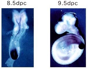
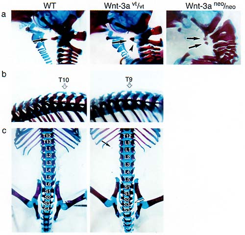
The research interest of this laboratory is to understand molecular mechanism of the vertebrate development. Particularly, roles of cell-to-cell signals, including members of Wnt and fibroblast growth factor (FGF) families, are characterized. Evidence indicated that each signal is involved in many aspects of the vertebrate development. For instance, we have revealed that Wnt-3a, a members of Wnt family, plays essential roles in a number of aspects of the mouse development, including somite development, neural crest formation and neural development. However, cellular and molecular mechanisms how a cell signaling molecule regulates these different events. Thus, we are focusing on precise functional analysis of cell-to-cell signals and identification of target genes induced by these signals. In addition, to reveal molecular networks in which these signals are involved, we are also trying genetical approach with the zebrafish. I Roles of Wnt signals in somite developmentThe Wnt family of genes that encode cysteine rich secreted proteins consists of at least 15 members in the vertabrate. It has already been shown that some of them are expressed and play important roles during gasturulation. For instance, Wnt-3a, a member of the Wnt family genes, is expressed in the primitive streak ectoderm during gastrulation and in the tailbud in later development of the mouse (Fig. 1). For dissecion of the complex developmental events regulated by Wnt-3a signaling in these regions, it is important to identify genes regulated by this signal. It has already been demonstrated that T (Brachyury) is a direct target of Wnt-3a in the anterior primitive ectoderm, which is fated to give rise to the paraxial mesoderm, suggesting that Wnt-3a modulates a balance between mesodermal and neural cell fates via T. To gain more insight into the role of Wnt-3a during gastrulation, we searched for potential target genes of Wnt-3a. We found that Wnt signaling stimulated the in vitro expression of cdx-1, and that Wnt-3a was required for full activation of cdx-1expression in the primitive ectoderm and tailbud in vivo. Moreover, Wnt-3a mutants displayed a defect in their anteroposterior patterning similar to that in the cdx-1 mutant mouse, in addition to another homeotic transformation that was not observed in cdx-1mutants (FIG. 2). These results suggest that cdx-1 is one of the mediator genes of Wnt-3a signaling in the anteroposterior patterning of the vertebrae and that Wnt-3a is also involved in a Cdx-1 independent process in anteroposterior patterning
II A roles of Wnt signaling in neural developmentIn the dorsal half of the spinal cord, three subclasses of interneurons called D1, D2 and D3 from the dorsal side. These interneuron subclasses are thought to be derived from distinct neural progenitor domains. Recent studies indicate that the roof plate is a source of inductive signals that control the generation of D1 and D2 classes of dorsal interneurons. Interestingly, Wnt-1 and Wnt-3a are expressed in largely overlapping regions within the central nervous system, predominantly in the roof plate from diencephalon to spinal cord. We show that mouse embryos lacking both Wnt1 and Wnt3a are indeed defective in determination of the dorsal interneurons. Generation of D1 and D2 classes of dorsal interneurons and their progenitors was markedly disrupted; this loss of the dorsal interneurons was compensated by a dorsal expansion D3 interneuron populations. Most importantly, expression of TGFb family members was not significantly affected in these mutant embryos. Moreover, we demonstrate that WNT3A can induce D1 and D2 class interneurons in the isolated intermediate region of the neural tube. Together, these observations clearly indicate that Wnt signaling has a critical role in the generation of dorsal interneurons specific D1 and D2 cell types. III Screening of target genes of Wnt signaling by an gene trap approachTo gain more insight into roles of Wnt signaling during embryogenesis, we searched for potential target genes of this signaling by an induction gene trap screening in mouse ES cells. In at least three ES cell clones among 794 clones screened, expression of beta-geo reporter genes was dramatically changed in response to the conditioned medium of Wnt-3a expressing cells. The expression analysis of the reporter genes in embryos generated from these ES cell clones revealed that the spatiotemporal expression patterns of these reporter genes were well correlated to those of several Wnt genes. These results suggested that an induction gene trap approach is effective for screening of target genes of Wnt signaling during embryogenesis. IV Roles of Fgf signals in bone developmentThe Fgf family of cell signaling molecules is composed of at least 22 members in the mouse. Although Fgf signaling has been implicated in the bone development, study on null mutant mice have not yet fully shown the role of this family in the skeletal development. As an attempt to identify a candidate Fgf gene essential for bone formation, we have examined the expression of mouse Fgf18gene using in situ hybridisation and demonstrated expression during calvarial and long bone development. Furthermore, to investigate the role of Fgf18 in vivo, we generated a null allele of the Fgf18 locus by homologous recombination in ES cells. In Fgf18-deficient mouse embryos generated by gene targeting, progress of the suture closure is delayed. Furthermore, terminal differentiation of the osteoblast is specifically delayed in developing calvaria and long bones. Proliferation of calvarial osteogenic mesenchymal cells is transiently decreased. On the other hand, the number of proliferating and differentiated chondrocytes is increased in the limbs. Fgf18 is thus essential for distinct aspects of cell proliferation and differentiation in bone formation. Taken together, Fgf18 plays essential roles in the osteogenesis and the chondrogenesis of the mammal and regulates differently cell proliferation and differentiation in these two processes.. V Screening of mutations affecting mesoderm development in zebrafishTo understand the molecular mechanism of mesoderm development in the tail bud, we have been screening mutations affecting mesoderm development in zebrafish. Publication List:Hino, S.-I., Kishida, S., Michiue, T., Fukui, A., Sakamoto, I., Takada, S., Asashima, M. & Kikuchi, A. (2001) Inhibition of the Wnt signaling pathway by Idax, a novel Dvl-binding protein. Mol. Cell. Biol., 21, 330-342. Ikeya, M. & Takada, S. (2001) Wnt-3a is required for somite specification along anteroposterior axis of the mouse embryo and regulates cdx-1 expression. Mech. Dev.103, 27-33. Jin, E.J., Erickson, C.A., Takada, S,. & Burrus, L.W. (2001) Wnt and bmp signaling govern lineage segregation of melanocytes in the avian embryo. Dev. Biol. 233, 22-37. Kawamura, Y., Kikuchi, A., Takada, R., Takada, S., Sudoh, S., Shibamoto, S., Yanagisawa K., & Komano, H. (2001) Inhibitory effect of presenilin 1 mutation in the Wnt signaling pathway---presenilin 1mutations enhance the phosphorylation of b-catenin--- Eur. J. Biochem. 268 3036-3041. Koshida, S., Shinya, M., Nikaido, M., Ueno, N., Schulte-Merker, S., Kuroiwa, A., & Takeda, H. (2002) Inhibition of BMP activity by the FGF signal promotes posterior neural development in zebrafish Dev. Biol. (in press). Mao, J., Wang, J., Liu, B., Pan, W., Farr, G.H., Flynn, C., Yuan, H., Takada, S., Kimelman, D., Li, L., & Wu, D. (2001) Low-density lipoprotein receptor-related protein-5 binds to axin and regulates the canonical Wnt signaling pathway. Mol. Cell7, 801-809. Muroyama, Y., Fujihara, M., Ikeya, M,. Kondoh,. H., & Takada, S.(2002) Wnt signaling plays an essential role in neuronal specification of the dorsal spinal cord. Genes Dev. 16, 548-55. Nomi M., Oishi I., Kani S., Suzuki H., Natsuda T.,Yoda A., Kitamura M., Itoh K., Takeuchi S Tkeda K., Akira S., Ikeya M., Takada S., & Mnami Y. (2001) Loss of mRor1 enhances the heart and skeletal abnormalities in mRor2-deficient mice: kinase. Mol Cell. Biol. 21, 8329-8335. Ohbayashi, N., Shibayama, M., Kurotaki, Y., Imanishi, M., Fujimori, T., Itoh, N., & Takada, S. (2002) Fgf18 is required for normal cell proliferation and differentiation during osteogenesis and chondrogeneses. Genes Dev. (in press) Shinya, M., Koshida, S., Sawada, A., Kuroiwa, A., & Takeda, H. (2001) Fgf signalling through MAPK cascade is required for development of the subpallial telencephalon in zebrafish embryos. Development128, 4153-4164. Yamane, T., Kunisada, T., Tsukamoto, H., Yamazaki, H., Niwa, H., Takada, S. & Hayashi, S.-I. (2001) Wnt signaling regulates hematopoiesis through stromal cells. J. Immunol.167, 765-772. |
|
Copyright(C) National Institute for Basic Biology (webmaster@nibb.ac.jp) |