DIVISION OF SPECIATION MECHANISMS I
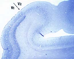
Our research goal is to understand mechanisms underlying evolution of the nervous system. In order to approach this question, we are studying the genes that are expressed in specific areas of the primate neocortex. Using differential display method, we have obtained genes that showed marked differences within the primate neocortex. Our second approach is to understand informational processing in the brain underlying learning behaviors with gene expression techniques. Here, we report our findings in the year of 2001. I. Genes expressed in specific areas of the neocortexThe neocortex is most evolved in mammals, particularly in primates, and thought to play the major role in higher functions of the brain. It is known to be divided into distinct functional and anatomical areas and has been a matter of debate what extent the area of the neocortex are genetically and environmentally determined. It is also puzzling why, during the evolution of mammals, the neocortex was most markedly expanded while the number of the genes in the mammal was little changed. To access these questions, we studied gene expression within different areas of the neocortex. 1) In collaboration with Professor Hiroyuki Nawa (Nigata university), we used the DNA macroarray technique to examine gene expression in the areas of human prefrontal, motor and visual cortexes. We found almost all the genes among 1088 genes examined showed only less than a factor of two in the difference of their expressions. Only one gene showed more than three fold difference and another one was between two and three fold difference within the three areas. These results suggest that the genes that are expressed among the different areas of the human neocortex are very similar. However, the question remained whether there are any genes that show marked difference within areas of neocortex. 2) In order to answer this question, we employed differential display methods and found at least two genes that indicated area specific expressions. i) One gene, designated occ1, is specifically expressed in the occipital cortex, particularly in V1 area, in the primate brain. Furthermore, the expression of occ1 turned out to be activity dependent, because, in the monocularly deprive-monkeys injected with TTX into one of the eyes, the expression of occ1 is markedly decreased in the ocular dominance columns of the primary visual cortex (V1). ii) The other gene that showed marked difference within the neocortex, is gdf7, a member of BMP/TGF-b family, which is specifically expressed in the motor cortex of the African green monkey. We are currently examining the detailed expression pattern of the gene. 3) We have also further isolated several area specific genes with RLCS (Restriction Landmark cDNA Scanning). II. Gene expression under audio-visual discrimination taskWe studied gene expression of c-Fos under audio-visual discrimination tasks in collaboration with professor Yoshio Sakurai (Kyoto University). We found that the visual and audio tasks enhanced the specific expression of c-Fos in the visual and audio cortices, respectively. Among the early visual and auditory pathways examined, c-Fos was specifically induced in the cortices but not in the earlier pathways, suggesting the neural modulation of the neocortex depending on the types of the tasks. Interestingly, the task-dependent Fos expression was only observed in excitatory neurons in the relevant sensory cortices. III CNTF is specifically expressed in the developing pineal glandsCNTF, a member of the IL-6 family, attracts quite attentions of developmental neuroscientists because it shows various effects on neurons and glial cells. The CNTF knockout mice, however, only indicates moderate motor neuron deficiency in the adult, but no apparent phenotype in the development. In order to explore the function of the IL-6 family, we extensively examined the expression of members of the family and their receptors and found the specific expression of CNTF in the embryonic pineal glands and eyes. Since sympathetic neurons are known to innervate pineal glands and be responsive to CNTF, our observation suggests that CNTF plays a role when sympathetic neurons innervate pineal glands.
Publication list:Original ArticlesFujiwara, T., Yamamori, T. and Akagawa, K. Suppression of transmitter release by Tat HPC-1/syntaxin 1A fusion protein. (2001) Biochim. Biophys. Acta1539, 225-232. Hanazawa, A., Mikamai, A., Angelika, P.S., Takenaka, O., Goto, S., Onishi, A., Koike, S., Yamamoi, T., Kato, k., Kondo, A., Suryobroto, B., Farajallah, A., Komatsu, H. (2001) Electroretinogram analysis of relative spectral sensitivity in genetically identified dichromatic macaques. Proc. Natl Acad. Sci. USA., 98, 8124-8127. Hata, K., Arakai, M. and Yamamori, T. CNTF is specifically expressed in the developing rat pineal gland and eyes. NeuroReport(in press) Karachot, L., Shirai, Y., Vigot, R., Yamamori, T. and Ito, M. Induction of long-term depression in cerebellar Purkinje cells requires a rapidly turned over protein. (2001) J. Neurophysiol. 86, 280-289. Onishi, A., Koike, S., Ida-Hosonuma, M., Imai, H., Shichida, Y., Takenaka, O., Hanazawa, A., Komatsu, H., Mikamai, A., Goto., S., Suryobroto, B., Farajallah, A., Varavudhi, P., Ekavhibata, C., Kitahara, K. and Yamamori, T. Variations in long and middle-wavelength-sensitive opsin loci in crab-eating monkeys. Vision Res. (in press) Sakata, S., Kitsukawa, T., Kaneko, T., Yamamori, T. and Sakurai, Y. Task-dependent and cell-type-specific Fos enhancement in rat sensory cortices during audio-visual discrimination. Eur. J. Neuroscience (in press) Tochitani, S., Liang, F., Watakabe, A., Hashikawa, T., and Yamamori, T. (2001) occ1 is preferentially expressed in the primary visual cortex in an activity-dependent manner: a pattern of gene expression related to the cytoarchitectonic area in adult macaue neocortex. Eur. J. Neurosci. 13, 297-307. Vigot, R., Batini, C., Kado, R.T. and Yamamori. T. Synaptic LTD in vivo recorded on the rat cerebellar cortex. Arch. Ital. Biol.(in press) Watakabe A. , Sugai T., Nakaya N., Wakabayashi K., Takahashi H., Yamamori T. and Nawa H. (2001) Similarity and Variation in Gene Expression Among Human Cerebral Cortical Subregions Revealed By DNA Macroarrays: Technical Consideration of RNA Expression Profiling from Postmortem Samples. Mol. Brain Res., 88, 74-82. Watakabe, A., Fujita, H., Hayashi, M. and Yamamori T. (2001) GDF7, a BMP/TGF beta family member, is enriched in the primary motor area of monkey neocortex. J. Neurochem., 76, 1455-1464. |
|
Copyright(C) National Institute for Basic Biology (webmaster@nibb.ac.jp) |