DIVISION OF BEHAVIOR AND NEUROBIOLOGY (ADJUNCT)
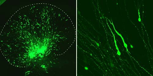
The vertebrate nervous system contains a large number of neurons that are connected to each other by specific axonal projections. We are interested in how the complex but precise neuronal network, which is indispensable for functioning of the nervous system, is constructed during development. The goal of our research in this division is to elucidate the cellular and molecular mechanisms underlying the neuronal network formation in vertebrates, with current focuses on the mechanisms of axon guidance and neuronal migration. I. Mechanism of Axonal GuidanceDuring development of the nervous system, growing axons are guided through specific pathways to correct targets. Our research interest focuses on the cellular and molecular mechanisms of axon guidance in the vertebrate nervous system. We are particularly interested in how guidance mechanisms operating in different phases of axonal growth are assembled to generate the complex but precise axonal wiring patterns in the brain. To address these issues, we are first trying to understand axonal wiring patterns during development with reference to the structural organization of the brain. Using whole-mounted preparations of embryonic rat brains, we will reveal whole axonal trajectories by labeling with neuronal tracers, such as DiI, or by immunohistochemical staining with antibodies against molecular markers expressed in a subset of neurons. We will next explore the axonal guidance mechanisms generating these wiring patterns. Following questions will be addressed. 1) What structures along the pathway have key roles in axonal guidance? 2) What guidance cues exist in these structures? Are these attractive cues or repulsive cues? Are these short-range cues or long-range cues? 3) What molecules are responsible for these cues? 4) How do multiple guidance mechanisms work in concert to generate specific wiring patterns? To answer these questions, we use neuroanatomical techniques, in vitro culture techniques including dissociated cell, explant and whole-mount cultures, and biochemical and molecular biological techniques. II. Mechanism of Neuronal MigrationA variety of neurons migrate from their birthplace to the position where they finally settle. Neuronal migration in the vertebrate central nervous system occurs both along the radial axis and along the tangential axis of the neural tube. Another interest of our research focuses on the cellular and molecular mechanisms underlying the radial and tangential neuronal migration. In many regions of the central nervous system, neurons migrate radially from the ventricular zone where they are born toward the pial surface. We are investigating the mechanisms of radial migration, using neurons in the neocortex as a model system. In addition to the radial migration, some neurons in the brain, such as interneurons in the forebrain, move tangentially for a long distance. We are also investigating how tangentially migrating neurons are guided for a long distance through specific routes to the final positions, using neurons that migrate from the rhombic lip at the dorsal rim of the hindbrain, such as cerebellar granule cells and precerebellar neurons. To address these issues, we developed in vitro culture systems that reconstruct migratory events occurring in vivo. We labeled migrating neurons with green fluorescent protein (GFP) by transplantation of small piece of explant taken from a transgenic rat expressing GFP or by introducing GFP cDNA into limited regions of the brain by electroporation. Slices of the brain or flat-mounted brain preparation are then cultured on permeable membrane filters. These culture systems enable us to analyze migration pattern of neurons in real time (Fig. 1). Using these in vitro culture systems, we are currently investigating the cellular and molecular mechanisms of neuronal migration.
Publication List:Imai, T., Tokunaga, A., Yoshida, T., Hashimoto, M., Mikoshiba, K., Weinmaster, G., Nakafuku, M., and Okano, H. (2001). The neural RNA-binding protein Musashi1 translationally regulates mammalian numb gene expression by interacting with its mRNA. Mol. Cell.Biol.21, 3888-900. Mizuguchi, R., Sugimori, M., Takebayashi, H., Kosako, H., Nagao, M., Yoshida, S., Nabeshima, Y., Shimamura, K., and Nakafuku, M. (2001). Combinatorial roles of olig2 and neurogenin2 in the coordinated induction of pan-neuronal and subtype-specific properties of motoneurons. Neuron31, 757-71. Nakashima, K., Takizawa, T., Ochiai, W., Yanagisawa, M., Hisatsune, T., Nakafuku, M., Miyazono, K., Kishimoto, T., Kageyama, R., and Taga, T. (2001). BMP2-mediated alteration in the developmental pathway of fetal mouse brain cells from neurogenesis to astrocytogenesis. Proc. Natl. Acad. Sci. USA98, 5868-73. Otsuka, T., Murakami, F., and Song, W. J. (2001). Excitatory postsynaptic potentials trigger a plateau potential in rat subthalamic neurons at hyperpolarized states. J. Neurophysiol.86, 1816-25. Sawamoto, K., Nakao, N., Kobayashi, K., Matsushita, N., Takahashi, H., Kakishita, K., Yamamoto, A., Yoshizaki, T., Terashima, T., Murakami, F., Itakura, T., and Okano, H. (2001). Visualization, direct isolation, and transplantation of midbrain dopaminergic neurons. Proc. Natl. Acad. Sci. USA98, 6423-8. Shirasaki, R., and Murakami, F. (2001). Crossing the floor plate triggers sharp turning of commissural axons. Develop. Biol.236, 99-108. Tashiro, Y., Miyahara, M., Shirasaki, R., Okabe, M., Heizmann, C. W., and Murakami, F. (2001). Local nonpermissive and oriented permissive cues guide vestibular axons to the cerebellum. Development128, 973-81. |
|
Copyright(C) National Institute for Basic Biology (webmaster@nibb.ac.jp) |