DIVISION OF MOLECULAR NEUROBIOLOGY
1 CREST, JST
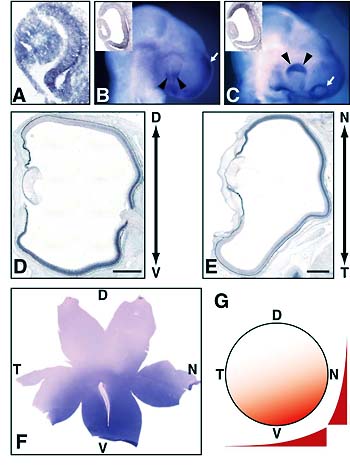
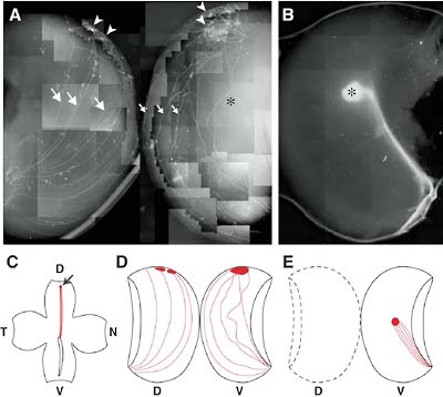
We have been studying the molecular and cellular mechanisms underlying the development and functioning of the vertebrate central nervous system. The scope of our interests encompasses regional specification in the retina, neuronal differentiation, cellular migration, path-finding and target recognition of axons, formation and refinement of specific synapses, and also various functions of the matured brain. I. Molecular mechanism of the retinotectal projectionTopographic maps are a fundamental feature of neural networks in the nervous system. Understanding the molecular mechanisms by which topographically ordered neuronal connections are established during development has long been a major challenge in developmental neurobiology. The retinotectal projection of lower vertebrates including birds has been used as a readily accessible model system. In this projection, the temporal (posterior) retina is connected to the rostral (anterior) part of the contralateral optic tectum, the nasal (anterior) retina to the caudal (posterior) tectum, and likewise the dorsal and ventral retina to the ventral and dorsal tectum, respectively. Thus, images received by the retina are precisely projected onto the tectum in a reversed manner. Since 1992, we have been devoting our efforts to searching for topographic molecules which show asymmetrical distribution in the embryonic chick retina. In the first-round screening using a cDNA subtractive hybridization technique, we identified two winged-helix transcriptional regulators, CBF-1 and CBF-2, expressed in the nasal and temporal retina, respectively. Furthermore, our misexpression experiments using a retroviral vector showed that these two transcription factors determine the regional specificity of the retinal ganglion cells, namely, the directed axonal projections to the appropriate tectal targets along the anteroposterior axis. To further search for topographic molecules in the embryonic retina, we next performed a large-scale screening using a new cDNA display system called Restriction Landmark cDNA Scanning (RLCS). With the assistance of a computer image-processing software, we successfully identified 33 molecules along the nasotemporal axis and 20 molecules along the dorsoventral axis, with various asymmetrical expression patterns in the developing retina. We have elucidated the primary structures of all these cDNA clones and examined their expression patterns during development. These included many novel molecules together with the known molecules: transcription factors (CBF-2, COUP-TFII, etc.), receptor proteins (EphA3, EphB3, etc.), secretory factors, intracellular proteins, and so on. Among them, we identified a novel retinoic acid (RA)-generating enzyme, RALDH-3, which is specifically expressed in the ventral region of the retina, together with a dorsal-specific enzyme RALDH-1. Furthermore, we recently identified a novel secretory protein, Ventroptin, which has BMP-4 neutralizing activity (Fig. 1). Ventroptin is expressed in the retina with a ventral high-dorsal low gradient at early stages. This expression pattern is complementary to that of BMP-4. At later stage (E6-8), a nasal high-temporal low gradient expression pattern is also detected. Ventroptin thus shows a double-gradient expression profile along the dorsoventral and the anteroposterior axes. Misexpression of Ventroptin altered expression patterns of several topographic genes: BMP-4, Tbx5 and cVax along the dorsoventral axis and ephrin A2 along the anteroposterior axis. Consistently, in these embryos, projection of the retinal ganglion cell axons to the tectum was also changed along the both axes (Fig. 2). The topographic retinotectal projection along the dorsoventral and the anteroposterior axes thus appears to be controlled not separately but in a highly concerted manner by Ventroptin. Currently, with respect to the identified topographic molecules, we are conducting over- and misexpression experiments using viral vectors and in ovo electroporation to elucidate their molecular functions. We expect that our studies will lead to elucidation of the molecular mechanism underlying the retinal patterning and topographic retinotectal projection, and ultimately to uncovering the basic principles for establishing complicated but extremely precise neural networks.
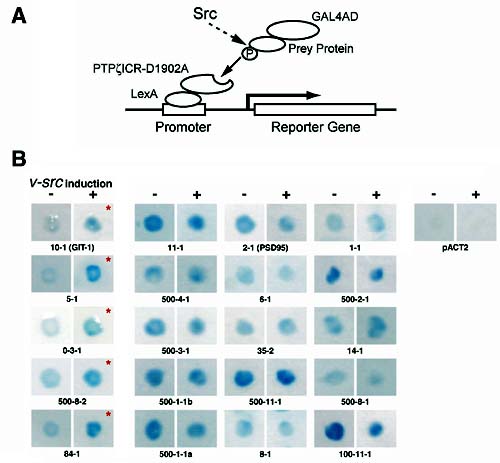
II. Functional roles of protein tyrosine phosphatase z and gProtein tyrosine phosphorylation plays crucial roles in various aspects of brain development and brain functions. In 1994, we found that PTPz/RPTPz, a nervous system-rich RPTP, is expressed as a chondroitin sulfate proteoglycan in the brain. The extracellular region of PTPz consists of a carbonic anhydrase-like domain, a fibronectin-type III-like domain and a serine-glycine-rich region, which is considered to be the chondroitin sulfate attachment region. There exist three splice variants of this molecule: a full-length transmembrane form (PTPz-A); a short transmembrane form (PTPz-B); and a soluble form (PTPz-S), which is also known as 6B4 proteoglycan/phosphacan. PTPz is expressed from the early developmental stage to the adulthood. This suggests that this gene plays variegated roles in the brain development and brain function. We began by searching for ligand molecules of this receptor. We found in 1996 that PTPz binds pleiotrophin/HB-GAM and midkine, closely related heparin-binding growth factors which share many biological activities. The chondroitin sulfate portion of PTPz is essential for the high affinity binding (Kd = ~0.25 nM) to these growth factors, and removal of chondroitin sulfate chains results in a marked decrease of binding affinity (Kd = ~13 nM). We further revealed that chondroitin sulfate interacts with Arg78 in Cluster I, one of the two heparin-binding sites in the C-terminal half domain of midkine. This is the first demonstration that chondroitin sulfate plays an important regulatory role in growth factor signaling. Next, we examined the roles of pleiotrophin/midkine-PTPz interaction in neuronal migration using the glass fiber assay and Boyden chamber cell migration assay. Pleiotrophin and midkine on the substratum stimulated migration of neurons in these assays. Experiments using various midkine mutants with various affinities for PTPz indicated that the strength of binding affinities and the neuronal migration-inducing activities are highly correlated. These results suggest that PTPz is involved in migration as a neuronal receptor for pleiotrophin and midkine. In order to reveal the intracellular signaling mechanism of PTPz, we performed yeast two-hybrid screening using the intracellular region of PTPz as bait. We found in 1999 that PTPz interacts with PSD-95/SAP90 family members, SAP102, PSD-95/SAP90 and SAP97/hDlg, which are concentrated in the central synapses mediating protein-protein interactions to form large synaptic macromolecular complexes. Here, the C-terminus of PTPz binds to PSD-95/SAP90 proteins through the second PDZ domain. This suggests that PTPz is involved in the regulation of synaptic function. However, PSD-95/SAP90 family members are not likely to be the substrate for PTPz because this family members are not tyrosine-phosphorylated. To identify the substrate molecules of PTPz, we have recently developed the yeast substrate-trapping system. This system is based on the yeast two-hybrid system with two essential modifications: conditional expression of v-src to tyrosine-phosphorylate the prey proteins and screening using a substrate-trap mutant of PTPz as bait. Using this system, we successfully isolated a number of candidate clones for substrate molecules or interacting molecules (Fig. 3). Among them, we first identified GIT1/Cat-1 as a PTPz substrate. It is known that Cat-1 regulates Pak, a serine threonine kinase which serves as a target for the small GTP-binding proteins, Cdc42 and Rac, and is implicated in a wide range of cellular events including the cell adhesion and cell morphological change. Pleiotrophin, PTPz and GIT1/Cat-1 might regulate the neuronal migration and neurite extension by controlling the Pak signaling pathway. We are continuing efforts to characterize the other candidate clones. To further study the physiological function of PTPzin vivo, we generated PTPz-deficient mice in which the PTPz gene was replaced with the LacZ gene in 1997. By investigating the expression of LacZ in heterozygous mutant mice, we demonstrated that neurons as well as astrocytes express PTPz in the central nervous system. We are currently studying the phenotype of PTPz-deficient mice biochemically, anatomically, physiologically and ethologically, and have already found abnormalities in behavior, learning and memory, etc.
III. Physiological roles of Nax sodium channelHuman Nav2.1, mouse Nav2.3 and rat NaG/SCL11 were cloned as a subfamily of voltage-gated sodium channel (NaCh). We found that these genes are species orthologs. This channel molecule is divergent from the previously cloned NaChs (Nav1.1-1.9) including the regions involved in activation, inactivation and ion selectivity. In the end of 2000, it was renamed Nax as a novel enigmatic member of sodium channels. To clarify the physiological function and cells expressing Nax sodium channels in vivo, we generated knock-out mice in which Nax channel gene was replaced with the LacZ or neo gene by gene targeting. Analysis of the targeted mice allowed us to identify Nax-producing cells by examining the lacZ expression. Besides in the lung, heart, dorsal root ganglia and Schwann cells in the peripheral nervous system, Nax was expressed in neurons and ependymal cells in restricted areas of the central nervous system, particularly in the circumventricular organs that are involved in body-fluid homeostasis (see Fig. 1 in the part of Center for Transgenic Animals and Plants). The null mutant mice showed markedly elevated c-fos expression in neurons in the subfornical organ and organum vasculosum laminae terminalis compared with wild-type animals. This suggests that these neurons are in a hyperactive state in the Nax-deficient mice. Moreover, the null mutants showed abnormal intakes of hypertonic saline. These findings suggest that the Nax sodium channel plays an important role in the central sensing of the body-fluid sodium level, and in regulation of salt intake behavior. We are currently examining differences in the electrophysiological property between the cells derived from the Nax-null mutant mouse and wild-type mouse to gain insight into the channel property of Nax.
Publication List:Kawachi, K., Fujikawa, A., Maeda, N. and Noda, M. (2001) Identification of GIT1/Cat-1 as a substrate molecule of protein tyrosine phosphatase z/b by the yeast substrate-trapping system. Proc. Natl. Acad. Sci. USA98, 6593-6598. Qi, M., Ikematsu, S., Maeda, N., Ichihara-Tanaka, K., Sakuma, S., Noda, M., Muramatsu, T. and Kadomatsu, K. (2001) Haptotactic migration by midkine: Invovement of protein-tyrosine phosphatase z, mitogen-acti-vated protein kinase and phosphatidylinositol 3-kinase. J.Biol. Chem., 276, 15868-15875. Sakuta, H., Suzuki, R., Takahashi, H., Kato, A., Shintani, T., Iemura, S., Yamamoto, T.S., Ueno, N. and Noda, M. (2001) Ventroptin: A BMP-4 antagonist expressed in a double-gradient pattern in the retina. Science293, 111-115. Shintani, T., Maeda, N. and Noda, M. (2001) Receptor-like protein tyrosine phosphatase g (RPTPg), but not PTPz/RPTPb, inhibits NGF-induced neurite outgrowth in PC12D cells. Dev. Neurosci., 23, 55-69. Sugawara, T., Tsurubuchi, Y., Agarwala, K. L., Ito, M. Fukuma, G., Mazaki-Miyazaki, E., Nagafuji, H., Noda, M., Imoto, K., Wada, K., Mitsudome, A., Kaneko, S., Montal, M., Nagata, K., Hirose, S. and Yamakawa, K. (2001) A missense mutation of the Na+ channel aII subunit gene Nav1.2 in a patient with febrile and afebril seizures causes channel dysfunction. Proc. Natl. Acad. Sci. USA98, 6384-6389. Thomaidou, D., Coquillat, D., Meintanis, S., Noda, M., Rougon, G. and Matsas, R. (2001) Soluble forms of NCAM and F3 neuronal cell adhesion molecules promote Schwann cell migration: identification of protein tyrosine phosphatase z/b as the putative F3 receptors on Schwann cells. J. Neurochem.78, 767-778. Zubair, M., Watanabe, E., Fukada, M. and Noda, M. (2002) Genetic labeling of specific axonal pathways in the mouse central nervous system. Eur. J. Neurosci., in press. Abstracts:Fujikawa, A., Watanabe, E., Sakaguchi, G., Katsuura, G., Hattori, S., Song, W.J., Noda, M. Dopmainergic dysfunction in the mice lacking the receptor tyrosine phosphatase z/RPTPb gene. In the abstracts of the 31th Society for Neuroscience of North America. 2001. Hiyama, T.Y., Watanabe, E., Yoshida, S., Noda, M. Nax channel is involved in monitoring extracelular sodium concentration. ibid. Kawachi, H., Fujikawa, A., Maeda, N. Noda, M. Identification of GIT1/Cat-1 as a substrate molecule of protein tyrosine phosphatase z/b by the yeast substrate-trapping system. ibid. Sakuta, H., Suzuki, R., Takahashi, H., Kato, A., Shintani, T., Iemura, S., Yamamoto,T.S., Ueno, N., Noda, M. Ventroptin: A novel BMP-4 antagonist expressed in a double-gradient pattern in the retina. ibid. . Suzuki, R., Shintani, T., Sakuta, H., Kato, A., Ohkawara, T., Osumi, N., Noda, M. Novel retinoid dehydrogenases expressed in the retina. ibid. Watanabe, E., Fujikawa, A., Matsunaga, H., Yasoshima, Y., Sako, N., Yamamoto, T., Noda, M. Nax channel is involved in control of salt intake behavior in CNS. ibid |
|
Copyright(C) National Institute for Basic Biology (webmaster@nibb.ac.jp) |