DIVISION OF REPRODUCTIVE BIOLOGY
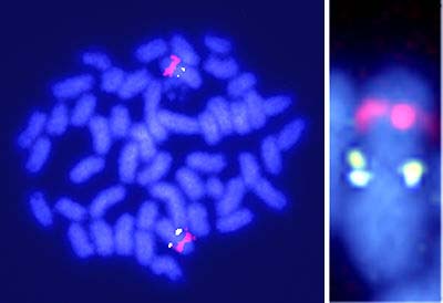
The pituitary-gonadal axis plays an important role in regulating gametogenesis in vertebrates. Gonadotropins typically act through the biosynthesis of gonadal steroid hormones which in turn mediate various stages of gametogenesis. Their effects are particularly profound in teleost fishes which provide several excellent models for investigating the basic hormonal mechanisms regulating gonadal sex differentiation and gametogenesis (spermatogenesis, sperm maturation, oocyte growth and oocyte maturation). Our research focuses on (1) the identification of steroidal mediators involved in gonadal sex differentiation and gametogenesis, and (2) the mechanisms of synthesis and action of these mediators. I. Sex-determining gene of medakaAlthough the sex-determining gene, Sry, has been identified in mammals, no comparable genes have been found in non-mammalian vertebrates. We used positional cloning to identify the sex-determining gene of medaka, Oryzias latipes. Chromosome walking using a BAC library and DNA markers derived from a congenic strain mapped the sex-determining region to a 530 kb stretch of the Y chromosome. We found a congenic XY female medaka lacking 250 kb of this region, further shortening the probable sex-determining region. Shotgun sequencing of this deleted region predicted 27 genes; however, only three were expressed in embryos during sexual differentiation. Furthermore, only one (PG17) of these three genes was present specifically on the Y chromosome (Fig. 1). We also found a naturally-occurring XY female. Offspring of this female inherited a Y chromosome that contained, but did not express, PG17 suggesting an important role for PG17 in testis development. Based on its homology with the DM gene family, Y chromosome specificity, and apparent role in testis development, PG17 was identified as the sex-determining gene of medaka.
II. Endocrine regulation of gonadal sex differentiationNile tilapia, Oreochromis niloticus, is an excellent example of the precise nature of steroidogenic actions during gonadal sex differentiation. In this fish, all genetic female (XX) or male (XY) broods can be obtained by artificial fertilization of normal eggs (XX) and sex-reversed, pseudo male sperm (XX), or normal eggs (XX) and super male sperm (YY), respectively. Fertilized eggs hatch after 4 days at 26°C. On the day of hatching, primordial germ cells (PGCs), are located in the outer layer of the lateral plate mesoderm around the hind gut. At 3 days post-hatching, PGCs are located in the gonadal anlagen after the formation of the coelomic cavity in the lateral plate mesoderm rather than through active migration. The gene vasaencodes a DEAD (Asp-Glu-Ala-Asp) family of putative RNA helicase and is present in the germ line of several animal species. Two isoforms of vasa mRNA and protein are present in tilapia. One (vas-s) lacks a part of the N-terminal region found in the other isoform (vas). Both isoforms are expressed in oocytes through the embryonic stage when PGCs localize in the lateral plate mesoderm. After PGCs localization in the gonadal anlagen, vas-s expression increases and vas expression becomes undetectable. Expression of both isoforms is observed again after morphological gonadal sex differentiation, irrespective of genotypic sex. In ovary, compared to vasexpression vas-s expression predominates throughout oogenesis. In testis, vas expression is predominant compared with vas-sduring spermatogenesis. These results indicate that relative expression of two vasa isoforms is dependent upon germ cell differentiation and sex. We have also generated medaka transgenic lines with green fluorescent protein (GFP) fluorescence controlled by the regulatory regions of the olvas gene in the germ cells. The intensity of GFP fluorescence increases dramatically in PGCs located in the ventrolateral region of the posterior intestine around stage 25 (the onset of brood circulation). Whole-mount in situ hybridization and monitoring of ectopically located cells by GFP fluorescence suggest that 1) the increase in zygotic olvas expression occurs after PGC specification and 2) PGCs can maintain their cell characteristics ectopically after stages 20-25. The GFP expression persists throughout the later stages in the mature ovary and testis. In tilapia, mitosis of germ cells begins around 10 days post-hatching in genetic females, but can not be confirmed until after sex differentiation in testes of genetic males. Steroid-producing cells in ovaries, but not testes, at the undifferentiated and differentiating stages express all of the steroidogenic enzymes required for estradiol-17b biosynthesis from cholesterol. Transcripts of estrogen receptors (ER) a and b first appear in both female and male gonads of fry 5-10 days post-hatching. These results strongly suggest that endogenous estrogens act as the natural inducers of ovarian differentiation in tilapia. This hypothesis is further supported by evidence of masculinization of genetic female tilapia by inhibition of estrogen synthesis using an inhibitor of cytochrome P450 aromatase. In contrast, the ability of steroid-producing cells to synthesize steroid hormones in the testes only appears at the time of testicular differentiation. Transcripts of androgen receptors are not present in gonads of genetic males during sex differentiation. We have isolated two DM (Doublesex/Mab-3 DNA-binding motif)-domain cDNAs from tilapia testis and ovary, named DMRT1 and DMO, respectively. DMRT1 is expressed only in Sertoli cells and DMO is detected only in oocytes by in situhybridization. The correlation between expression of DMRT1 and testicular differentiation of both normal XY-male and sex reversed XX-males suggest that DMRT1 is a candidate testis-determining gene in tilapia. In contrast, abundant DMO expression in pre- and early vitellogenic oocytes in XX- and sex reversed XY-females indicates a relationship between DMO and oocyte growth. III. Endocrine regulation of spermatogenesisSpermatogenesis is an extended process of differentiation and maturation of germ cells resulting in haploid spermatozoa. Using an organ culture system for eel testes consisting of spermatogonia and inactive somatic cells, we have shown that the hormonal regulation of spermatogenesis in eel testes involves the gonadotropin stimulation of Leydig cells to produce 11-ketotestosterone (11-KT), a potent androgen in fish. In turn, 11-KT activates Sertoli cells to stimulate the production of activin B. Addition of recombinant eel activin B to the culture medium induced proliferation of spermatogonia, producing late type B spermatogonia, within 15 days in the same manner as did 11-KT. cDNAs encoding two androgen receptors (ARa and ARb) have been cloned, for the first time in any vertebrates, from eel and tilapia testes. In situ hybridization reveals that although both AR mRNAs are present in eel testes prior to human chorionic gonadotropin (hCG) injection, only ARa transcripts increase during hCG-induced spermatogenesis suggesting that ARa and ARb play different roles in spermatogenesis. Activin B binds to activin type I and II receptors on spermatogonia to stimulate denovo synthesis of G1/S cyclins and CDKs leading to the initiation of mitosis. Interestingly, cyclin A1 transcripts are first detected in primary spermatocytes during hCG-induced spermatogenesiss in eel testes suggesting an important role for cyclin A1 in the progression to meiosis of male germ cells. IV. Endocrine regulation of oocyte maturationMeiotic maturation of fish oocytes is induced by the action of maturation-inducing hormone (MIH). 17a, 20b-dihydroxy-4-pregnen-3-one (17a,20b-DP) has been identified as the MIHs of several fish species. The interaction of two ovarian follilce cell layers, the thecal and granulosa cell layers, is required for the synthesis of 17a,20b-DP. The theal layer produces 17a-hydroxy- progesterone that is converted to 17a,20b-DP in granulosa cells by the action of 20b-hydroxysteroid dehydrogenase (20b-HSD). The preovulatory surge of LH-like gonadotropin is responsible for the rapid expression of 20b-HSD mRNA transcripts in granulosa cells during oocyte maturation. In tilapia, Northern blot and RT-PCR analyses reveal that the expression of 20b-HSD in ovarian follicles is not detectable in postvitellogenic follicles. A distinct expression is evident at the day of spawning. In vitro incubation of postvitellogenic follicles with hCG induced the expression of 20b-HSD mRNA within 1-2 hrs, followed by final oocyte maturation of oocytes. Actinomycin D completely blocks both hCG-induced 20b-HSD expression and final oocyte maturation, indicating the involvement and transcriptional regulation of 20b-HSD in final maturation. 17a,20b-DP induces oocyte maturation by acting on a pertussis toxin-sensitive G-protein-coupled membrane receptor. The early steps of l7a,20b-DP action involve the formation of downstream mediator of this steroid, the maturation-promoting factor or metaphase-promoting factor (MPF) consisting of cdc2 kinase and cyclin B. 17a,20b-DP induces oocytes to synthesize cyclin B which activates a preexisting 35-kDa cdc2 kinase via phosphorylation of its threonine 161 by a threonine kinase (MO15), thus producing the 34 kDa active cdc2. Polyadenylation of cyclin B mRNA is involved in 17a,20b-DP-induced initiation of cyclin B mRNA translation. The Mos/MAPK pathway is not essential for initiating goldfish oocyte maturation despite its general function as a cytostatic factor (CSF). Upon egg activation, MPF is inactivated by degradation of cyclin B. We showed that the 26S proteasome initiates cyclin B degradation through the first cut of its NH2 terminus at lysine 57. Publication List:Ikeuchi, T., Todo, T., Kobayashi, T. and Nagahama, Y. (2001) Two subtypes of androgen and progestogen receptors in fish testes. Comp. Physiol. Biochem. Part B129, 449-455. Ikeuchi, T., Todo, T., Kobayashi, T. and Nagahama, Y. A novel progestogen receptor subtype in the Japanese eel, Anguilla japonica. FEBS Letters(in press). Imahara, J., Tokumoto, M., Nagahama, Y., Ishikawa, K. and Tokumoto, T. (2001) Reconstitution of sperm nuclei of zebrafish, Danio rerio, in Xenopus egg extracts. Marine Biotechnology3, 53-57. Kitano, T., Takamune, K., Nagahama, Y. and Abe, S. (2001) Role of P450 aromatase in gonadal sex differentiation in Japanese flounder (Paralichthys olivaceus). Environmental Sciences, 8, 1-11. Kobayashi, T., Kajiura-Kobayashi, H. and Nagahama, Y. Two isoforms of vasa homologs in a teleost fish: their differential expression during germ cell differentiation. Mech. Develop.(in press). Lee, Y.H., Du, J.L., Yueh, W.S., Lin, B . Y., Huang, J. D., Lee, C. Y., Lee, M. F., Lau, E. L., Lee, F. Y., Morrey, C., Nagahama, Y. and Chang, C. F. (2001) Sex change in the protandrous black porgy, Acanthopagrus schlegeli: A review in gonadal development, estradiol, estrogen receptor, aromatase activity and gonadotropin. J. Exp. Zool.290, 715-726. Liu, S.J., Govoroun, M., D'Cotta, H., Ricordel, M.J., Lareyre, J.J., McMeel, O.M., Smith, T., Nagahama, Y. and Guiguen, Y (2001) Expression of cytochrome P450b (11b-hydroxylase) genes during gonadal sex differentiation and spermatogenesis of rainbow trout, Oncorhynchus mykiss. J. Steroid Biochem. Mol. Biol.75, 291-298. Morrey, C.E., Nagahama, Y. and Grau, E. G. Terminal phase males stimulate ovarian function and inhibit sex change in the protogynous wrasse Thalassoma duperrey. Zool. Sci. (in press). Matsuda, M. and Nagahama, Y. Positional cloning of the sex-determining region of medaka using a Y congenic starain. Aqua Genome (in press). Matsuda, M., Kawato, N., Asakawa, S., Shimizu, N., Nagahama, Y., Hamaguchi, S., Sakaizumi, M. and Hori, H. (2001) Construction of a BAC library derived from the inbred Hd-rR strains of the teleost fish, Oryzias latipes. Gene Genetic Systems76, 61-63. Matsuyama, M., Sasaki, A., Nakagawa, K., Kobayashi, T., Nagahama, Y. and Chuda, H. (2001) Maturation- inducing hormone of the tiger puffer, Takifugu rubripes (Tetraodontidae, Teleostei): biosynthesis of steroids by the ovaries and the relative effectiveness of steroid metabolites for germinal vesicle breakdown in vitro. Zool. Sci.18, 225-234. Nakahata, S., Katsu, Y., Mita, K., Inoue, K., Nagahama, Y. and Yamashita, M. (2001) Biochemical identification of Xenopus Pumilio as a sequence-specific cyclin B1 mRNA-binding protein that physiologically interacts with a Nanos homolog, Xcat-2, and a cytoplasmic polyadenylation element-binding protein. J. Biol. Chem.276, 20945-20953. Nakahata, S., Mita, K., Katsu, Y., Nagahama, Y. and Yamashita, M. (2001) Immunological detection and characterization of poly(A) polymerase, poly(A)-binding protein and cytoplasmic polyadenylation element- binding protein in goldfish and Xenopusoocytes. Zool. Sci.18, 337-343. Nakamura, M., Kobayashi, T., Yoshiura, Y. and Nagahama, Y. Masculinizing all-female tilapia by administrating aromatase inhibitor. Mol. Reprod. Develop. (in press). Oba, Y., Hirai, T., Yoshiura, Y., Kobayashi, T. and Nagahama, Y. (2001) Fish gonadotropin and thyrotropin receptors: The evolution of glycoprotein hormone receptors in vertebrates. Comp. Physiol. Biochem. Part B129, 441-448. Ohta, K., Rhaman, M.A., Chuda, H., Yoshikuni, M., Nagahama, Y. and Matsuyama, M. Maturation-inducing hormone and it's membrane receptor in gonads of Japanese yellowtail, Seriola quenqueradiata. Fisheries Science (in press). Tanaka, M., Kinosita, M. and Nagahama, Y. (2001). Establishment of medaka (Oryzias latipes) transgenic lines with the expression of GFP fluorescence exclusively in germline cells: a useful model to monitor germline cells in a live vertebrate. Proc. Natl. Acad. Sci. USA98, 2544-2549. Tanaka, M., Nakajin, S., Kobayashi, D., Fukada, S., Guan, G. J., Todo, T., Senthilkumaran, B. and Nagahama, Y. . Teleost ovarian carbonyl reductase-like 20b-hydroxysteroid dehydrogenase: Potential role in the production of maturation-inducing hormone in final maturation of oocytes. Biol. Reprod.(in press). Tokumoto, M., Nagahama, Y. and Tokumoto, T. Molecular cloning of cDNA encoding a polypeptide chain elongation factor 1a from goldfish (Carassius auratus). DNA Sequence (in press). Tokumoto, M., Nagahama, Y. and Tokumoto, T. A major substrate for MPF: cDNA cloning and expression of polypeptide chain elongation factor 1gfrom goldfish (Carassius auratus). DNA Sequence (in press). Yamaguchi, A. and Nagahama, Y. Structure and protein compositions of the nuclear matirix prepared from germinal vesicles of goldfish (Carassius auratus) oocytes. Mol. Reprod. Develop. (in press). Yamaguchi, A. and Nagahama, Y. Somatic lamins in germinal vesicles of goldfish vitellogenic oocytes. Cell Structure and Function(in press). |
|
Copyright(C) National Institute for Basic Biology (webmaster@nibb.ac.jp) |