National Insitute for Basic Biology  


DIVISION OF MOLECULAR NEUROBIOLOGY


Professor:
Masaharu Noda
Research Associates:
Nobuaki Maeda
Shinji Hirano
Masahito Yamagata
Graduate Students:
Hiroki Hamanaka
Junichi Yuasa
Takafumi Shintani
Taeko Nishiwaki
Visiting Fellow:
Haruyuki Matsunaga *
Technical Staff:
Tomoko Mori
Shigemi Ohsugi
Masae Mizoguchi
(* from Environmental Health Science Laboratory, Sumitomo Chemical Co., Ltd.)

Our efforts have been devoted to studying molecular and cellular mechanisms which underlie the development of the vertebrate central nervous system. We are seeking for the molecules and structures that regulate various cellular events requisite for the brain morphogenesis, such as generation of neuroblasts, their migration to form the laminar structure and various nuclei, the elongation and path-finding of neural processes, and the formation and refinement of specific connections between neurons. The research has been conducted using various techniques, including molecular biology (e.g. cDNA cloning, site-directed mutagenesis), biochemistry (protein, carbohydrate), monoclonal antibodies, neuroanatomy, cell and organotypic culture, and embryo manipulation (classical embryology, gene transfer with viral vectors, and gene targeting).


I. Topographical and laminar connection in the chick retinotectal system

Neural connection in the vertebrate brain is selective in two ways, which we refer to as topographical and laminar. Topographical specificity determines the orderly maps of connectivity, in which presynaptic neurons arrayed along spatial axes project to corresponding arrays of target cells in the target area. By contrast, laminar specificity determines the local circuitry, in which presynaptic neurons form synapses on the particular cells/portions of the particular target layer/cell's surface. However, we still have no knowledge about the crucial factors that determine the development of these two classes of neural connection. We have chosen to study a retinotectal system in chickens which have many experimental advantages.

Most intensively studied in this system has been the retinotopic map, where retinal ganglion cells from various parts of the retina project topographically onto the tectal surface. For example, retinal axons from the temporal (posterior) retina connect to the anterior part of the tectum. Many works, since the formulation of 'chemoaffinity theory' by Roger Sperry, have supported the involvement of gradient molecules in this topographically organized connection. In positing this hypothesis, we performed subtractive screening to isolate the cDNAs that is graded along the anterio-posterior axis in the retina. Recently, we have found several known and novel molecules, which are selectively expressed in the nasal or temporal half region of the retina. Starting with these molecules, we hope to understand the molecular mechanism by which retinotectal topographical connection is established.

Less well studied, but comparatively striking, is a laminar selectivity of retinotectal connection in the orthogonal direction. Retinal axons enter the tectum through the most superficial of its 15 different laminae. Once axons reach defined loci on the tectal surface, they branch inwards and make connections in only 3-4 of the 15 Iaminae. Moreover, each axon confines most of its synapses to one of these 'retinoreceptive' laminae, and neurochemically-distinguishable subsets of the ganglion cells connect to distinct laminae to establish a functional network (Yamagata et al. submitted). The laminar selectivity in this system may require an axon to recognize particular target cells (cellular specificity) and particular portions of the target cell's surface (subcellular specificity). To identify the cellular and molecular mechanism underlying these synaptic specificities, we are taking two interrelated approaches. First, we are trying to establish culture systems to analyze the recognition cues between presynaptic axons and their target cells in vitro. The second approach is to use molecular techniques, particularly by developing useful probes. We are now preparing new monoclonal antibodies that mark the subsets of laminae or cells, eventually to identify the molecules that guide the lamina-specific connection. To understand the function of these molecules, we will employ a variety of gene transfer techniques, including retroviral and adenoviral vectors.


II. Proteoglycan and brain development

Proteoglycans are a family of proteins bearing sulfated glycosaminoglycans, which bind many extracellular matrix components and growth factors through their core protein and glycosaminoglycan portions. We have been interested in the functional roles of the brain-specific proteoglycans in the development of the nervous tissue because they are the major extracellular matrix components in the tissue.

Previously, we identified a phosphate-buffered saline-soluble chondroitin sulfate proteoglycan with a 300-kDa core protein (6B4 proteoglycan) using a monoclonal antibody (MAb6B4). Immunohistochemical analysis of the adult rat hindbrain showed that 6B4 proteoglycan is expressed in fairly restricted areas such as pontine nuclei and lateral reticular nucleus. Developmental studies of the rat hindbrain indicated that the expression of 6B4 proteoglycan is spatiotemporally correlated with the circuit formation of the mossy fiber system. In the embryonal rat cerebral cortex, in contrast, 6B4 proteoglycan is expressed on the radial glial fiber, a scaffold for neuronal migration.

Recently, we cloned cDNAs encoding this proteoglycan from a ƒÉgt11 rat whole brain cDNA library. Nucleotide sequence analysis of the isolated cDNA clones revealed that 6B4 proteoglycan is highly homologous to human receptorlike protein tyrosine phosphatase (PTPase), PTPƒÄ (also known as RPTPß). In parallel, Maurel et al. reported a cDNA encoding a chondroitin sulfate proteoglycan termed phosphacan, from rat brain, and it turned out that 6B4 proteoglycan is identical to phosphacan. The cDNA analysis including our own revealed that 6B4 proteoglycan/phosphacan is an alternatively spliced extracellular variant of PTPƒÄ. We then attempted to identify proteoglycan-type PTPases in the rat brain, where many types of proteoglycans and PTPases are known to be present.

Membrane-bound proteoglycan fractions were obtained from the postnuclear membrane preparation of the 8-day-old rat brain by DEAE ion exchange chromatography and CsCl density gradient centrifugation. The isolated proteoglycan fraction contained a high PTPase activity with typical PTPase characteristics. Protein renaturation experiments from SDS gels demonstrated that chondroitin sulfate proteoglycans with a 380- and 170-kDa core proteins carried the PTPase activity. The proteoglycan with 380-kDa core protein was identified as PTPĀ with the specific antibodies. The PTPase with a 170-kDa core protein did not cross-react with the antibodies against PTPĀ, suggesting that these two are not closely related structurally. These findings show that early postnatal rat brain indeed contain multiple proteoglycan-type PTPases.

Proteoglycans may play crucial roles in cell adhesion, motility, growth and differentiation through the process of binding to various extracellular matrix molecules and growth factors. On the other hand, the level of protein tyrosine phosphorylation is regulated by cellular interactions with these molecules through the modulation of protein tyrosine kinase and PTPase activities. Our findings that some membrane-bound PTPases are proteoglycans would explain the functional overlap between the two classes of molecules.

Fig.1

Fig. 1
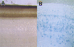
A) Lamina-specific projection of retinal axons in the chick optic tectum. The anterogradely-labeled retinal terminals (brown) make synapses in some restricted laminae. B) Recombinant adenovirus-mediated gene transfer into the chick optic tectum. Adenovirus carrying lacZ (E. coli ß-galactosidase) was injected into the mesencephalic ventricle. After one week, many cells in various laminae express the introduced gene (blue).


Publication List:

Maeda, N., Hamanaka, H., Shintani, T., Nishiwaki, T. and Noda, M. (1994) Multiple receptor-like protein tyrosine phosphatases in the form of chondroitin sulfate proteoglycan. FEBS Lett. 354, 67-70.

Maeda, N., Hamanaka, H., Oohira, A. and Noda, M. (1995) Purification, characterization and developmental expression of a brain-specific chondroitin sulfate proteoglycan, 6B4 proteoglycan/phosphacan. Neuroscience. in press.

Oohira, A., Matsui, F., Watanabe, E., Kushima, Y. and Maeda, N. (1994) Developmentally regulated expression of a brain specific species of chondroitin sulfate proteoglycan, neurocan, identified with a monoclonal antibody 1G2 in the rat cerebrum. Neuroscience. 60, 145-157.