9.
How to transform Physcomitrella patens
9.1
PEG-mediated transformation
Yuji Hiwatashi and Mitsuyasu
Hasebe
Transformation of Physcomitrella patens has been performed with the polyethylene glycol (PEG)-mediated DNA introduction to protoplasts, with the particle bombardment to live cells (see 9.2), and with the agrobacterium-mediated DNA introduction to protonemata or protoplasts (see 9.5). The PEG-mediated DNA transfer method is the most efficient and feasible for homologous recombination mediated-gene targeting. DNA bombardment usually provides unstable transformants without recombination into Physcomitrella genome and is mainly used for transient assay. Agrobacterium-mediated method is used for a specific purpose; such as high-throughput screening (see 9.5).
1. Preparation of protonemata for
transformation
(1)
Inoculate a piece of protonemata with tweezers to BCDATG medium and cultivate
for three weeks. The original protonemata are usually composed of chloronemata
and caulonemata. We will make chloronema-rich protonemata by the following
procedures, because transformation rates of chloronemata are higher than those
of caulonemata.
(2)
After each inoculated protonemata forms a colony after three weeks, move three
colonies to a test tube with 25 mL sterile water and chopped with a Polytron homogenizer
(Kinematica, AG, Lucerne, Switzerland) for 10 sec at the minimum speed of the
Polytron PT2100 with a DA2120/2 generator shaft or at speed level 4 for the
Polytron model K with a PTA20TS generator shaft.
(3)
Spread 2.5 ml of the solution of chopped protonemata on a pretreated cellophane
sheet layered on BCDATG medium (see 2.2) in a 9-cm Petri dish. Incubate the
inoculated Petri dish at 25°C under continuous white light (at an intensity of
30-80 µmol m-2 s-1) for 7 days without sealing the plate.
(4) Collect all protonemata on
the plate, chop with the above conditions, spread, and cultivate as (3) for
three times and you can get chloronema-rich protonemata.
(5) Collect the chloronema-rich
protonemata of (4), chop with the above conditions, spread, and cultivate under
the same conditions for 3 to 5 days instead of 7 days. The regeneration rate of protoplasts is
critical for transformation efficiency. Protonemata grown for 3 to 5 days after
spreading should be used for protoplast isolation. Older protonemata cause dramatically
low transformation efficiency.
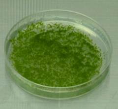
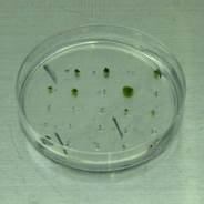
Protonemata
cultivated on a cellophane-overlaid plate for 5 days after chopping
2. Plasmid preparation
1. Culture E. coli harboring plasmid DNA.
2. Prepare plasmids using QIAGEN Hi-Speed
Plasmid Midi Kit or Promega Wizard Mini-prep Kit or other similar kit.
3. Digest the plasmid DNA with an appropriate
restriction enzyme to linearize.
4. Purify the digested plasmid with
phenol/chloroform extraction and precipitate by adding 1/10th the
volume of 3M NaOAc (pH5.2) and 2 volumes of ethanol.
5. Dissolve the precipitated plasmid with
TE, adjusting at the concentration of 0.2~1.0 µg/µl.
6. Store at –30˚C.
* If you use PCR amplified DNA
fragments, PCR products should be ethanol-precipitated with 1/10 volume of 3M
NaOAc (pH5.2) and resuspend in TE.
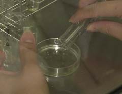
3. Transformation
1. (Day 1) Culture protonemata after
chopping. Grow them at 25˚C for 4 days (or 3 to 5 days).
2. (Day 4) Chop propagated protonemata
again and grow them at 25˚C for 4 days on a new plate.
3. (Day 8~9) Transformation followed by incubation
of transformed protoplasts in a top agar for 3 to 5 days.
4. (Day 12) Transfer the top agar
containing regenerating protoplasts onto a selection plate supplemented with
antibiotics. Cultivate for more than 3 weeks on the selection medium.
5. (Day 43; 3 weeks after TF) Transfer a fragment
of a colony grown onto the 1st selection plate to an antibiotic-free
plate. Incubate for more than one week.
6. (Day 50; 4 week after TF) Transfer a fragment
of a colony grown on the non-selection plate to a selection medium supplemented
with antibiotics. Incubate for more than one week.
7. (Day 57; 5 weeks after TF) Select each
survived colony on the selection medium as a stable transformant.
The First day
[Materials]
・ 200 ml flask x 1 (autoclave)
・ 300 ml flask x 2 (autoclave)
・ 10 ml pipette x 2 (autoclave)
・ 50 ml centrifuge tube x 2 (autoclave)
・ Funnel with a sheet of 50 µm nylon mesh
(autoclave)
・ Tweezers x 1 (autoclave)
・ Pretreated cellophane (autoclave: see
2.2)
・ Yellow tips for P200 pipetteman and
blue tips for P1000 pipetteman (autoclave)
・ 0.22 µm syringe-driven filter unit and
10 ml syringe x 2
・ 0.45 µm syringe-driven filter unit and 50
ml syringe x 1
・ Neubauer hemacytometer (Becton
Dickinson no. 424011)
・ Water bath x 2
・ Centrifuge
・ 50 ml round-bottom tube (Falcon, Iwaki)
(sterile)
・ 15 ml round-bottom tube (Falcon, Iwaki)
(sterile)
・ 15 ml conical tube (Falcon 2057)
(sterile)
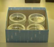
・ 6-cm Petri dish (sterile)
・ Parafilm
[Solution]
・ 500 ml 8% (w/v) mannitol solution
(autoclave)
・ 2 g PEG6000 in a 10 ml vial and a small
stir bar (autoclave)
・ 1% (w/v) MES (pH5.6)
・
・ 100 ml protoplast liquid medium
(autoclave)
|
H2O |
90 ml |
|
Stock A* |
1 ml |
|
Stock B* |
1 ml |
|
Stock C* |
0.1 ml |
|
5 g/l Ammonium tartrate |
1 ml (final concentration 50 mg/l) |
|
Mannitol |
6.6 g (final concentration 6.6%) |
|
Glucose |
0.5 g |
|
|
Fill up to 100 ml with H2O |
Stock medium
500 ml 8% (w/v) Mannitol
1 M Ca(NO3)2
Solution
1 M MgCl2 Solution
Driselase (available from Sigma)
All of the following stock media are
stored at 4˚C.
Stock A (x 100) DO NOT autoclave
|
Ca(NO3)2 4H2O |
118 g (0.5 M) |
|
FeSO4 7H2O |
1.25 g (4.5 mM) |
|
|
Fill up to 1000 ml with H2O |
Stock B (x 100) Autoclave
|
MgSO4 7H2O |
25 g (0.1 mM) |
|
|
Fill up to 1000 ml with H2O |
Stock C (x 100) Autoclave
|
KH2PO4 |
25 g (1.84 mM) |
|
|
Adjust to pH6.5 with 4M KOH |
|
|
Fill up to 1000 ml with H2O |
[Procedures]
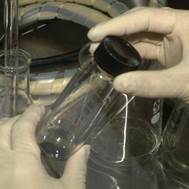
1. Add 0.5 g of Driselase in a 50 ml
conical tube and add 25 ml of 8% mannitol solution. Mix well.
2. Add 1 ml of 1 M Ca(NO3)2
and 100 µl of 1 M Tris-HCl (pH8.0) into 9 ml of 8% (w/v) mannitol solution and
mix. Filter the solution with a 0.22 µm filter.
3. (Preparation of PEG/T) Add 5 ml of the
filtered solution to the autoclaved PEG (see above). Dissolve PEG completely.
This solution is called PEG/T.
4. (Preparation of MMM) Mix 910 mg of
mannitol, 0.15 ml of MgCl2, 1 ml of 1% MES (pH5.6) and 8.85 ml of H2O.
Filter the solution with a 0.22 µm filter. This solution is called MMM.
5. Switch-on two water baths at 45˚C and at
20˚C.
6. Centrifuge the driselase solution at 4,000
rpm for 5 min.
7. Transfer the supernatant to a 50 ml
syringe with a 0.45-µm filter-unit and filtrate into a 50 ml centrifuge tube.
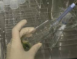
8. Transfer the propagated chloronema-rich
protonemata (1 to 2 g fresh weight, which are collected from 10 plates) into
the Driselase solution with sterile tweezers. Since protonemata growing close
to the edge of a Petri dish may be contaminated with bacteria, we collect protonemata
more than 1 cm inside from the edge. Then incubate at 25˚C for 30 min. Mix
gently every 5 min.
9. Filtrate the protonemata through 50-µm nylon-mesh.
10. Centrifuge the filtrated protoplasts at 1,000
rpm (180 x g) for 2 min at room temperature (22°C), and resuspend gently in 40
ml of 8% (w/v) mannitol. Repeat this washing procedure twice.
11. Count the number of resuspended
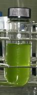
protoplasts with hemacytometer and re-suspend at 1.6 x 10⁶ ml-1
in MMM solution.
MMM (ml) = the number of protoplasts
per square (large nine squares) x 104 cell/ml x 40 ml / (1.6 x 10⁶
cell/ml)
12. Add 30 µl of plasmid DNA solution into a
15 ml round-bottom tube (Falcon 2057). Then, add 300 µl of the protoplast
suspension and 300 µl of PEG/T solution to the
tube. Mix gently.
13. Incubate the tubes containing the
transformation mixture in the 45˚C water bath for 5 min, then in the 20˚C water
bath for 10 min.
14. Dilute the transformation mixture with
adding 5 aliquots of 300 µl protoplast liquid medium at 3 min intervals and
then 5 aliquots of 1 ml of protoplast liquid medium at 3 min intervals.
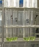
15. Pour the diluted protoplast solution into
a 6 cm Petri dish, seal it with Parafilm, and incubate at 25˚C overnight in
darkness.
[Key points]
・ Resuspend the protoplasts in the MMM
solution with gently pipetting if protoplasts aggregate at the bottom of the
centrifuge tube.
・ Use a round-bottom tube (for example, 14 ml, 17 mm x 100
mm, BD Falcon tube, no. 352057, Becton Dickinson, Franklin Lakes, NJ) to mix the solution more gently
The second day
[Materials]
・ 10 ml disposable pipette (sterile) or
white tips for P5000 pipetteman (autoclave)
・ Tweezers x 1 (autoclave)
・ Cellophane (autoclave) –
Quality of cellophane is different depending on companies. For regular
cellophane, it is better to be pre-treated as follows. If you use P-5 cellophane
(Futamura Chemical Co., Ltd, Nagoya), we can use
without EDTA treatment. However, other cellophanes may be better to be treated
as follows:
1) Cut cellophanes to a little
bit smaller than the size of a 9 cm-petri dish.
2) Place cellophane in a 500
ml beaker (usually ** seats) and then add 5 mM EDTA solution (pH8.0).
Autoclave.
3) Wash with MilliQ water
several times. Add MilliQ water in the beaker. Autoclave.
4) Place cellophane in a glass
petri dish and add MilliQ water, then autoclave.
When we use P-5 Cellophane
1)
Cut cellophanes to a little bit smaller than the size of a 9 cm-petri dish.
2)
Place cellophane in a 500 ml beaker and then autoclave.
3)
Place cellophane in a glass petri dish and add MilliQ water, then autoclave
・ 15 ml conical tube (Falcon)
・ Centrifuge
・ Surgical tape
[Solution]
・ PRM/T (200 ml); Autoclave and store at
45°C
|
H2O |
180 ml |
|
Stock B* |
2 ml |
|
Stock C* |
2 ml |
|
Stock D* |
2 ml |
|
Alternative TES |
0.2 ml |
|
500mM Ammonium tartrate |
2 ml (final concentration 5 mM) |
|
Mannitol |
16 g (final concentration 8%) |
|
CaCl2 2H2O |
0.29 g (final concentration 10 mM) |
|
Agar (Sigma: A6924)* |
1.6 g (final concentration 0.8%) |
|
|
Fill up to 200 ml with H2O |
* Agar provided by NAKARAI is not
appropriate.
・ PRM/B (1,000 ml); Autoclave and pour to
9 cm petri dishes. Dishes are preserved at room temperature.
|
H2O |
900 ml |
|
Stock B* |
10 ml |
|
Stock C* |
10 ml |
|
Stock D* |
10 ml |
|
Alternative TES |
1 ml |
|
500 mM Ammonium tartrate |
10 ml (final concentration 5 mM) |
|
Mannitol |
60 g (final concentration 6%) |
|
CaCl2 2H2O |
1.47 g (final concentration 10 mM) |
|
Agar (Sigma, A6924, Nacalai Tesque: cat. no. 01028-85) |
8 g (final concentration 0.8%) |
|
|
Fill up to 1000 ml with H2O |
* Stock
medium
All stock media are stored at 4˚C.
Stock D should be used within 2-3 months, before iron precipitates.
Stock B (x 100) Autoclave
|
MgSO4 7H2O |
25 g (0.1 mM) |
|
|
Fill up to 1000 ml with H2O |
Stock C (x 100) Autoclave
|
KH2PO4 |
25 g (1.84 mM) |
|
|
Adjust to pH6.5 with 4M KOH |
|
|
Fill up to 1000 ml with H2O |
Stock D (x 100) DO NOT autoclave
|
KNO3 |
|
|
FeSO4 7H2O |
1.25 g (4.5 mM) |
|
|
Fill up to 1000 ml with H2O |
[Procedures]
1. Overlay a sheet of pretreated cellophane
on a 9 cm-dish containing PRM/B medium.
2. Transfer the protoplast
suspension into a 15 ml polypropylene conical tube (15 ml, 17 mm x 120 mm, BD
Falcon tube, no. 352196, Becton Dickinson, Franklin Lakes, NJ)
with a pipette and centrifuge at 1,000 rpm (180 x g) for 2min at room
temperature (22°C).
3. Discard the supernatant and add 8 ml of a
PRM/T medium stored at 45°C. Re-suspend the protoplasts by pipetting.
4. Pour 2 ml of the protoplast suspension on
the pretreated cellophane layered on a PRM/B medium.
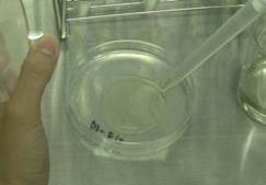
5. Seal the Petri dish using a surgical
tape. Incubate the plate at 25˚C for 3 days under continuous white light with a
light flux of 30 - 80 µmol m-2 s-1. We usually use 50 µmol m-2 s-1.
The third to fifth days
Incubate
the plates.
The sixth day
[Materials]
・ Tweezers x 2 (autoclave)
・ Surgical tape
[Solution]
・ Selection medium (BCDAT supplemented
with adequate antibiotics)
|
H2O |
900 ml |
|
Stock B |
10 ml |
|
Stock C |
10 ml |
|
Stock D |
10 ml |
|
Alternative TES |
1 ml |
|
500mM Ammonium tartrate |
10ml (final concentration 5 mM) |
|
50mM CaCl2 2H2O (if powder) |
20 ml (final concentration 1 mM) (0.15 g) |
|
Agar (Sigma; A6924, Nacalai Tesque: cat. no. 01028-85) |
8 g (final concentration 0.8%) |
|
|
Fill up to 100 ml with H2O |
After autoclaving, allow to cool to ~50˚C
and add appropriate antibiotics into the medium. Store at 4˚C.
Antibiotics used for selection
a) Geneticin (G418)
50 mg/mL solution is available from
Invitrogen (no. 10131-035).
Use at the final concentration of 20
mg/l.
b) Hygromycin B
50 mg/mL solution is available from
Invitrogen (no. 10687-010).
Use at the final concentration of 20
mg/l in the medium.
c) Zeocin
100 mg/mL solution is available from
Invitrogen (no. R250-01).
Use at the final concentration of 50
mg/l in the medium.
!NOTE!
Zeocin is light sensitive. Store zeocin solution and plates or medium containing zeocin in the dark.
d) Blasticidin S
One g of Blasticidin S, Hydrochloride (Funakoshi Co. Ltd KK-400) is dissolved in sterile water in a laminar cabinet, dissolved at 45°C for 10 min, pour 1 mL each into sterile 1.5 ml tubes. This stock solution is preserved at 4°C and use within 6 months. The stock solution is dissolved at 45°C for 10 min and used at the final concentration of 100 mg/L.
e) Nourseothricin/LEXSY NTC
LEXSY NTC (Jena Bioscience, no. AB-101S) is the trade name for the natural product nourseothricin (a complex of the streptothricins F and D) produced by Streptomyces noursei .
Use at the final concentration of 75 mg/l in the medium.
1. Insert tweezers under the cellophane and
transfer the protoplasts all together by lifting the cellophane disc with
tweezers to a BCDAT medium supplemented with adequate antibiotics.
2. Seal the Petri dish with a surgical tape.
Incubate it at 25˚C for 3~4 weeks under continuous white light.
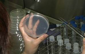
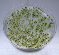 A plate after 3-week selection
[Key points]
・ Zeocin is light sensitive and zeocin
plates have to be preserved in the dark before use.
Transfer to drug-free medium (after an
incubation of 3-4 weeks on selection medium)
[Materials]
・ Tweezers (autoclave)
・ BCDAT plate
・ Surgical tape
[Procedure]
1. Transfer each colony under antibiotic
selection to antibiotics-free BCDAT medium. Tweezers are sterilized with 70% alcohol
and fired each time after transfer.
2. Culture at 25˚C for 1 week under
continuous light.
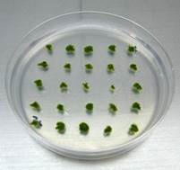 One-week colonies on antibiotics-free
BCDAT medium after inoculation
One-week colonies on antibiotics-free
BCDAT medium after inoculation
[Key points]
・ Transfer each colony one by one to a
selection medium to avoid generating a chimeric colony.
・ Incubate the plate for more than one week.
Transfer to selection medium (after an
incubation of 1 week on drug-free medium)
[Materials]
・ Tweezers (autoclave)
・ BCDAT plate containing adequate
antibiotics
・ Surgical tape
[Procedure]
1. After the incubation for one week without
antibiotics, transfer a small part of each protonemal colony to the BCDAT
medium supplemented with adequate antibiotics. A small part of a colony should
be transferred to remove chimeric colonies.
2. Seal the plates with a surgical tape and
incubate for one week.
3. Stable transformants grow on the plate.
[Key points]
・ Transferred protonemata should be
collected from the leading edge of a colony to prevent collecting chimeric
clones. Do not transfer a whole colony or gametophores.
・ When using plates containing zeocin, ensure
they are freshly prepared.
・ Some kinds of disruptants may show
reduced growth. Do not miss positive transformants even if growth of the colony
is reduced.
・ PEG-mediated transformation generates polyploids by protoplast fusion. Morphology of polyploid protonemata is not distinguished from that of wild type. DNA amount of transformants should be examined using flow cytometry (see chapter 8. Flow Cytometry Analysis).