10.2 How to observe the cellular
localization of a fluorescent protein
Takashi Murata and Yoshikatsu
Sato
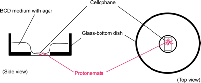
For analyzing cellular localization of a protein fused to a fluorescent protein, we need to observe protonemata without damage under good optical conditions. For this purpose, protonemata are cultured along the glass surface of a glass-bottomed dish (IWAKI 3910-039). The thickness of the agar medium is minimized to obtain good optical conditions. Additionally, having a large mass of agar medium connected with the thin layer of the medium, in which the protonemata grow, allows continuous nourishment and growth of the gametophyte.
Methods
1.
Pour 2 ml of BCD medium onto the plastic part of the glass-bottom dish and
allow to set for 30 min.
2. Pour 65 µl of BCD medium on the glass central
region of the dish.
3.
A small colony of protonemata is inoculated into the
glass-bottom dish with medium, and then overlaid with cellophane. After 3 or 4
days of culture, protonemata grow parallel to the
bottom of the dish.
Observation
should be performed by an inverted microscope.