OFFICE OF DIRECTOR
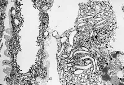
Mechanisms determining the outline shape of the adult lepidopteran wingsKODAMA, Ryuji Wings of the lepidopteran insects (butterflies and moths) develop from the wing imaginal disc, which is a hollow sac made of simple epithelium. When the pupariation is completed, the wing, which was hidden inside the body wall of the larvae, is exposed on the surface of the pupa, which gradually turns into the adult wing. The outline shape of the adult wing is often different from that of the pupal wing. This difference is brought about by the programmed cell death of the marginal area of the pupal wing, while the internal area develops as adult wing blade. The marginal dying area is called the degeneration region and the internal area is called the differentiation region, hereafter. The cell deaths in the degeneration region proceeds very rapidly and completes in a half to one day period in Pieris rapae or several other species examined. It was shown that the dying cells in the regeneration region have two characteristics common with the apoptotic cell death in mammalian cells. These are i) the presence of apoptotic bodies, which are heavily condensed cells or their fragments engulfed by other cells or macrophages, shown by transmission electron microscopy and ii) the presence of conspicuous accumulation of fragmented DNA evidenced by the TUNEL histological staining (Kodama, R. et al., Roux's Arch. Dev. Biol. 204, 418-426, 1995). The cells in the degeneration region are actively engulfed by the macrophages in the cavity beneath the wing epithelium. Moreover, the macrophages seem to be concentrated beneath the degeneration region by the strong adhesion between basal surfaces of the dorsal and ventral epithelium in the differentiation region. By injecting the india ink or ferritin solution to the body cavity of the pupa, we have confirmed that this adhesion is tight enough to exclude the macrophages from the differentiation region, because the injected probes was found mostly concentrated in the degeneration region when observed several minutes later (Yoshida, A. (Biohistory Research Hall) and Kodama, R., unpublished). A collaborative work with the laboratory of Dr. K. Watanabe (Hiroshima University) concerns mostly on the development of trachea and tracheole pattern in the swallow tail butterflies. Trachea and trcheoles are both important in delivering air into the wing and their pattern coincide with that of the boundary of degeneration and differentiation zones at the distal end of the wing. According to the observations, the pattern formation of wing epithelium is often dependent on tracheal and tracheole patterns. Basic research on the development of tracheal pattern formation is being done through the scanning electron microscopy and the bright field light microscopy of the fixed or fresh specimens to describe the exact pathway and the time course of the formation of elaborate pattern of trachea and tracheoles and to establish the cytological and developmental relationship between the formation of tracheal pattern and epithelial cell pattern, such as scale cell pattern. The figure depicts how the tracheoles protrude from the primary trachea at the pre-pupa stage (Fig. 1). The tracheoles are arranged with even spaces within the wing lumen and may closely related with the scale cell pattern formation. In collaboration with other developmental biologists, a database of cellular behaviors, which are essential steps of morphogenesis of multicellular organisms, is deviced (S3-P7, 14th ICDB, Kyoto, 2001). An instance of the database is basically made up of (i) the subject cell, (ii) the input the cell receives, and (iii) the output the cell excecutes. With the aid of methodologies in the field of information science and computer science, automatization of database collection is planned. This database will be important in incorporating rapidly increasing knowledge in developmental biology in highly reusable form even by non-expert researches of this filed.
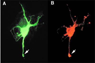
Protein palmitoylation and its role in neural developmental at embryogenesisUENO, Kohji Protein palmitoylase modifies specific cysteine residues of signalling proteins such as G proteins and G protein-coupled receptors with palmitate via thioester linkages. This modification is thought to be important in the regulation of signal transduction. We have previously found that protein palmitoylase is expressed in neural cells during mouse embryogenesis. Immunocytochemical analyses revealed that growth cone-associated protein (GAP)-43 and Go, which is one of G proteins, were mainly localized in growth cones and the cell body of cultured primary neural cells. Fig. 2 shows the localization of protein palmitoylase and GAP-43 in a neural cell. Cysteine residues in the amino terminal regions of GAP-43 and Go are palmitoylated. Furthermore an inhibitor of protein palmitoylase reduces the axonal growth of cultured neurons. From these findings, we speculated that the localization of the palmitoylated proteins in growth cones are critical for the development of axons. In this study, we are attempting to elucidate the mechanism that determines the localization of palmitoylated proteins in growth cones. For this analysis, we have established a method to chemically modify the amino terminal region of a synthetic GAP-43 peptide with palmitate or other fatty acids via a thioester linkage. Using these acylated peptides, we plan to analyze the mechanism that regulates the localization of palmitoylated peptide in growth cones.
|
|
Copyright(C) National Institute for Basic Biology (webmaster@nibb.ac.jp) |