NATIONAL INSITUTE FOR BASIC BIOLOGY  


National Institute for Basic Biology

DIVISION OF CELL FUSION

(Adjunct)


Professor:
Issei Mabuchi
Associate Professor:
Hiroshi Abe
Research Associate:
Hirotaka Fujimoto


Cytokinesis in animal and some primitive eukaryotic cells is achieved by the progressive contraction of the cleavage furrow. The cleavage furrow contains a contractile apparatus, called the contractile ring, which is composed of a bundle of actin filaments that lies in the furrow cortex beneath the plasma membrane. It has been established that the contractile ring contracts as the result of interaction between actin filaments and myosin. However, little is known about process of its formation, mechanism that controls its formation, protein constituents, and its ultrastructure.The goal of our research is to solve these problems and thereby clarify the molecular mechanism of cytokinesis. For this purpose, we use three kinds of cells, namely, sea urchin egg, Xenopus egg, and the fission yeast Schizosaccharomyces pombe.

S. pombe is an excellent system to investigate the changes in the actin cytoskeleton during cell cycle since F-actin patches, F-actin cables and F-actin ring are only visible structures in the cell (Fig. 1). The F-actin ring is considered to correspond to the contractile ring in animal cells. It is formed during anaphase in this organism. In order to obtain basic features of the actin cytoskeleton in S. pombe, we investigated subcellular localization and interactions of actin and two actin cytoskeleton-related proteins, Cdc8 tropomyosin and actin-related protein 3 (Arp3), using specific antibodies and by gene disruption. Actin was localized to medial microfilamentous ring in the region of the septum during cytokinesis and to cortical patches by immunoelectron microscopy. F-actin cables were detected throughout the cell cycle by fluorescent staining with Bodipy-phallacidin. The cables were often linked to the patches and to the medial F-actin ring during its formation. Tropomyosin was localized to the F-actin ring and the cables. It was also distributed in the cell as patches, although co-localization with F-actin was not frequent. In cdc8ts mutant cells, F-actin cables were not observed although the F-actin patches were detected and cell polarity was maintained. These observations suggest that the F-actin cables may be involved in the formation of the F-actin ring, and that tropomyosin plays an important role in organizing both the F-actin ring and the F-actin cable, but is not involved in the F-actin patch formation or maintenance of cell polarity.

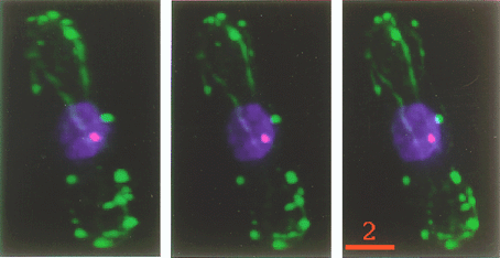
Fig. 1
3-D images of an interphase S. pombe cell. Each image is rotated by 12 degree from the neighbor. Green, F-actin. Blue, DNA. Red, spindle pole body. Bar, 2 mm.


Binding of Arp3 to actin was revealed by immunoprecipitation as well as by DNaseI column chromatography. Arp3 seemed to form a complex with several proteins in the cell extracts, as previously reported for other organisms. Arp3 was found to be concentrated in the medial region of the cell from early anaphase to late cytokinesis, however, it was not localized on the F-actin ring. Following arp3 gene disruption, F-actin patches were delocalized throughout the cell and cells did not undergo polarized growth, suggesting that Arp3 influences the proper localization of the actin patches in the cell and thereby controls the polarized growth of the cell.

Next we investigated the role of myosin in cytokinesis of S. pombe. Recently, it has been shown by Kitayama et al. (J. Cell Biol. 137: 1309-1319, 1997) that Myo2, a type II myosin heavy chain plays a role in the F-actin ring formation in this organism. We found another myosin II called Myo3. Myo3 is the same protein as Myp2 reported independently by Bezanilla et al. (Mol. Biol. Cell, 8: 2693-2705, 1997). Overexpression of Myo3 in the cell leads to formation of aberrant F-actin ring, F-actin cable, and septum. We knocked out the myosin genes in S. pombe. Since Myo3 is not essential, but Myo2 is essential for its growth, we made a myo2myo3 null strain containing pREP81-myo2 to control the expression of myo2. After shut off the myo2, no F-actin ring formation occurs during mitosis. Therefore, type-II myosin is necessary in the formation of the contractile ring in S. pombe.

We also concentrate our study on function of actin-regulatory proteins, including ADF/cofilin family proteins, during cytokinesis using Xenopus eggs and embryos. We found that ADF/cofilin family proteins are essential for cytokinesis (Abe, Obinata, Minamide, and Bamburg, J. Cell Biol. 132: 871-875, 1996). Recent studies revealed that ADF/cofilin accelerates turnover of actin filaments both in vitro and in vivo. Most recently, we found a novel actin-regulatory protein which induces disassembly of actin filaments cooperatively with ADF/cofilin. cDNA analysis revealed that this protein is a Xenopus homologue of yeast actin interacting protein 1 (AIP1). Thus, we designated this protein as Xenopus AIP1 (XAIP1). Purified XAIP1 itself binds to pure actin filaments to some extent, but it induces a rapid, drastic disassembly of actin filaments associated with cofilin. Microinjection of this protein into Xenopus embryos arrested development by the resulting actin cytoskeletal disorder. XAIP1 represents the first case of a protein cooperatively disassembling actin filaments with ADF/cofilin family proteins.


Publication List:
Hirata, D., Nakano, K., Fukui, M., Takenaka, H., Miyakawa, T. and Mabuchi, I. (1998). Genes that cause aberrant cell morphology by overexpression in fission yeast: a role of a small GTP-binding protein Rho2 in cell morphogenesis. J. Cell Sci. 111, 149-159.
Arai, R., Nakano, K. and Mabchi, I. (1998). Subcellular localization of actin, tropomyosin and actin-related protein 3 (Arp3) in Schizosaccharomyces pombe. Eur. J. Cell Biol. 76, 288-295.
Nishimura, Y., Nakano, K. and Mabuchi, I. (1998). Localization of Rho GTPase in sea urchin eggs. FEBS Lett. 441, 121-126.


webmaster@nibb.ac.jp
Last Modified: 12:00, May 28, 1999