NATIONAL INSITUTE FOR BASIC BIOLOGY  


National Institute for Basic Biology

DIVISION OF BEHAVIOR AND NEUROBIOLOGY

(Adjunct)


Professor:
Masatoshi Takeichi
Research Associate:
Akinao Nose
Kazuaki Tatei
Postdoctoral Fellow:
Emiko Shishido 1
Takako Isshiki 2
Graduate Students:
Hiroki Taniguchi
Takeshi Umemiya (from Kyoto University)


How individual nerve cells find and recognize their targets during development is one of the central issues in modern biology. The aim of our division is to elucidate the molecular mechanism of axon guidance and target recognition by using the simple and highly accessible neuromuscular system of Drosophila.

The musculature of Drosophila embryos consists of 30 identifiable muscle fibers per hemisegment. Each muscle fiber is innervated by a few motoneurons in a highly stereotypic manner. The high degree of precision and previous cellular manipulations of neuromuscular connectivity suggest the presence of recognition molecules on the surface of specific muscle fibers which guide the growth cones of motoneurons. We have previously isolated several enhancer trap lines that express the reporter gene b-galactosidase (b-gal) in small subsets of muscle fibers prior to innervation. By molecularly characterizing these lines, we are trying to identify genes that play roles in the specification of the muscles and neuromuscular connectivity. Previous studies showed that two of the lines are insertions in the connectin and Toll genes, that encode cell recognition molecules which belong to the leucine-rich repeat (LRR) family. We have been studying the function of these genes, and also characterizing other lines by molecular and genetic analysis.


I. Connectin can function as an attractive target recognition molecule.

Connectin is expressed on a subset of muscle fibers (primarily lateral muscles) and on the axons, growth cones of the motoneurons which innervate these muscles (primarily SNa motoneurons) and on several associated glial cells. When coupled with its ability to mediate homophilic cell adhesion in vitro, these results led to the suggestion that Connectin functions as an attractive signal for SNa pathfinding and targeting.

To study the role of Connectin in vivo, we ectopically expressed Connectin on all muscles by using MHC (myosin heavy chain) promoter (MHC-connectin) in the P-element mediated transformants. In MHC-connectin, SNa nerves were observed to send extra axon branches that form ectopic nerve endings on muscles 12, muscles they would never innervate in wild type. Furthermore, the ectopic innervation on muscle 12 was dependent on the Connectin expression on SNa. These results showed that Connectin functions as an attractive and homophilic guidance molecule for SNa in vivo.


II. Capricious, a novel LRR cell surface molecule expressed on subsets of neurons and muscles.

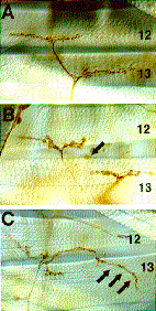
We have been conducting molecular and genetic analysis of other muscle enhancer trap lines. One of them, P750 expresses b-gal in subsets of neurons and muscles, including the RP5 motoneuron and its target, muscle 12. The cDNA cloning and sequencing revealed that the corresponding gene, termed capricious (caps), encodes a novel transmembrane protein that belongs to LRR family. It is interesting that three of the five muscle enhancer trap lines that we have thus far characterized contain LRRs. Within the LRR family, Caps protein was found to be most related to the product of the Drosophila tartan gene that have been implicated in neural and muscular development. We found that in the loss-of-function mutants of caps, the synaptic arborization pattern on muscle 12, a caps-positive muscle, was abnormal (Fig.1A, B). The nerve terminal failed to stabilize on muscle 12, and instead extended to and arborized on the adjacent muscle, muscle 13. Ectopic expression of caps in all muscles by GAL4-UAS system also resulted in aberrant synapse formation on muscle 12 (Fig. 1C). Like in the loss-of-function alleles, the muscle 12 terminal axon branch often formed collaterals that turned back and innervated muscle 13. These results suggest that Caps is involved in neuromuscular target recognition and/or stabilizaion of the synapses.

Fig. 1
Neuromuscular defects associated with loss-of-function and ectopic expression of caps. Body wall fillet of wild-type (A), caps loss-of-function (B) and caps misexpressing (C) 3rd instar larvae, stained with MAb 22C10 to visualize motor nerve projection and synaptic endings. Ectopic synapses formed by muscle 12 motoneurons are indicated by arrows.


III. msh, a homeobox containing gene essential for neural and muscular development.

Another line rH96 was found to be a P-element insertion in the muscle segment homeobox (msh) gene, that was previously cloned as a homeobox containing gene. By generating and analyzing both loss-of-function and gain-of-function (ectopic expression) mutants, we showed that msh is essential for neural and muscular development. During CNS development, msh is specifically expressed in the dorsal neuroectoderm and subsequently in many neuroblasts and their progenies derived from this region. We found that the loss of msh results in the failure of the proper differentiation of many neural and glial progenies derived from the dorsal neuroectoderm. Conversely, ectopic expression of msh in the entire neuroectoderm severely disrupts the formation of midline structure and differentiation of neuroblasts located in the ventral neuroectoderm. These results suggest that msh plays crucial roles in the dorso-ventral (DV) specification of the CNS. The vertebrate homologues of msh, Msxs are also know to be expressed in the dorsal portion of the spinal cord. Our work on msh raises a possibility that this family of genes may play a conserved role during DV patterning of the CNS.


IV. M-spondin and G-spondin: a novel gene family of secreted molecules

By molecularly characterizing another enhancer trap line, AN34 which is also expressed in a subset of muscles and neurons, we identified a novel secreted protein, termed M-spondin, that is highly homologous to rat F-spondin. F-spondin is a secreted molecule expressed at high levels in the floor plate and has been shown to promote neural cell adhesion and neurite extension in vitro. We found three regions that are highly conserved between M-spondin and F-spondin. One of them is a known repeating motif called thrombospondin type I repeats (TSRs). The other two domains (termed FS1 and FS2) are novel conserved sequences that we identified. By using PCR, we cloned two more genes that share similar overall structure with M-spondin and F-spondin in that they possessed FS1, FS2 and one to six TSRs. The identification of these genes thus defines a novel gene family of secreted protein with potential roles in cell adhesion. One of the newly cloned genes, termed G-spondin, is expressed in a subset of glia that sit along the longitudinal axon tracts in the CNS. The specific expression pattern of G-spondin suggests that it may play a role in the guidance of specific axons.


Publication List:
Oda, H., Uemura, T. and Takeichi, M. (1997) Phenotypic analysis of null mutants for DE-cadherin and Armadillo in Drosophila ovaries reveals distinct aspects of their functions in cell adhesion and cytoskeletal organization. Genes to Cells 2, 29-40.
Inoue, T., Chisaka, O., Matsunami, H. and Takeichi, M. (1997) Cadherin-6 expression transiently delineates specific rhombomeres, other neural tube subdivisions and neural crest subpopulations in mouse embryos. Develop. Biol. 183, 183-194.
Fushimi, D., Arndt, K., Takeichi, M and Redies, C. (1997) Cloning and expression analysis of cadherin-10 in the CNS of the chicken embryos. Develop. Dynamics 209, 1-17.
Nose, A., Umeda, T. and Takeichi, M. (1997) Neuromuscular target recognition by a homophilc interaction of Connectin cell adhesion molecules in Drosophila. Development 124, 1433-1441.
Radice, G.L., Rayburn, H., Matsunami, H., Knudsen, K.A., Takeichi, M. and Hynes, R.O. (1997) Developmental defects in mouse embryos lacking N-cadherin. Develop. Biol., 181, 64-78.
Umemiya, T., Takeichi, M. and Nose, A. (1997) M-spondin, a novel ECM protein highly homologous to vertebrate F-spondin, is localized at the muscle attachment sites in the Drosophila embryos. Develop. Biol. 186, 165-176.
Nakagawa, S. and Takeichi, M. (1997) N-cadherin is crucial for heart formation in the chick embryo. Develop. Growth Differ. 39, 451-455.
Iwai, Y., Usui, T., Hirano, S., Steward, R., Takeichi, M. and Uemura. T. (1997) Axon patterning requires DN-cadherin, a novel neuronal adhesion receptor, in the Drosophila embryonic CNS. Neuron 19, 77-89.
Isshiki, T., Takeichi, M. and Nose, A. (1997) The role of the msh homeobox gene during neurogenesis: implication for the dorsoventral specification of the neuroectoderm. Development 124, 3099-3109.
Suzuki, S.C., Inoue, T., Kimura, Y., Tanaka, T. and Takeichi, M. (1997) Neuronal circuits are subdivided by differential expression of type-II classic cadherins in postnatal mouse brains. Mol. Cell. Neuroscience 9, 433-447.
Higashijima, S., Nose, A., Eguchi, G., Hotta, Y. and Okamoto, H. (1997) Mindin/F-spondin family: novel ECM proteins expressed in the zebrafish embryonic axis. Develop. Biol., 192, 211-227.


webmaster@nibb.ac.jp
Last Modified: 12:00, May 28, 1998