NATIONAL INSITUTE FOR BASIC BIOLOGY  


National Institute for Basic Biology

DIVISION OF MORPHOGNESIS


Professor:
Naoto Ueno
Associate Professor:
Hiroshi Shibuya
Research Associates:
Makoto Nakamura
Makoto Mochii
Graduate Students:
Hideyuki Nagaso (Hokkaido University)
Masataka Nikaido (Hokkaido University)
Yoshihiro Takatsu (Hokkaido University)
Michiru Nishita (Hokkaido University)
Katsura Sugawara (The Graduate
University for Advanced Studies)
Satoru Yoshida (The Graduate
University for Advanced Studies)
Shinichi Ohki (Hokkaido University)
Yoshinori Tomoyasu (Hokkaido University)
Visiting Scientists:
Shinichi Higashijima (PRESTO, STA)
Kiyokazu Morita (Hokkaido University)
Fumihiko Hamada (Osaka University)
Technical Staffs:
Chiyo Takagi
Takamasa Yamamoto
Shunichiro Iemura
Yuko Takahashi


Our laboratory joined the NIBB in May of 1997 from Hokkaido University. We have been studying about the roles of growth factors in early embryogenesis.

Early developmental processes are regulated by a number of endocrine and paracrine factors that mediate cell-to-cell interactions. Particularly, polypeptide growth factors are believed to be essential for the regulation of cell proliferation and differentiation in morphogenesis during embryogenesis. One of the intriguing features of polypeptide growth factors is that their functions are well conserved in animal species. This allows us to study developmental mechanisms governed by growth factors using multiple animal models such as mouse, Xenopus, Drosophila, and C. elegans. We focus on a group of growth factors, the TGF-b superfamily and investigate molecular mechanisms for their actions in early embryogenesis. Our study involves biochemistry, embryology, molecular biology and genetics.


I. The role of follistatin in neural induction

It had been shown that bone morphogenetic protein BMP is a key regulator of mesoderm patterning by inducing ventral mesoderm in early Xenopus embryo. BMP also has an essential role in neural tissue formation. Presumptive ectoderm of Xenopus embryo gives rise to epidermis unless it is exposed to endogenous factors(s) from dorsal mesoderm or exogenous stimuli such as neural inducers. Therefore, it was believed that epidermis is a ground (default) state and neural tissue is induced by the neural inducers. However, it has been demonstrated by us and other groups that BMP specifies epidermis and inhibits neural fate. This anti-neural activity of BMP was later found to be conserved in animal species, from fly to mammals. More recently, it turned out that neural inducers chordin and noggin are BMP-binding proteins which inhibit BMP to interact with its receptor. These observations led to a model that not epidermal but neural fate is the default state. Follistatin, an activin-binding protein that inhibits activin activity had also been shown to act as a neural inducer. Because activin itself has no inhibitory action to neural induction, we questioned about the mechanism by which follistatin induces neural fate. Does follistatin mediate neural inducing signal through its own receptor yet unknown? Or, does it inhibit the neural inhibitor BMP as do chordin and noggin?

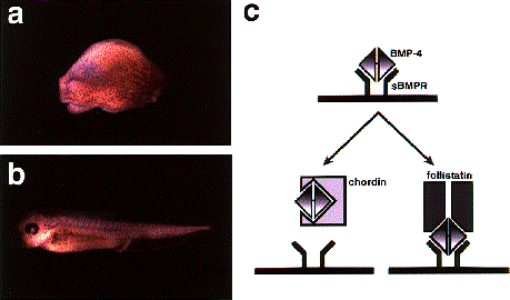
To address this problem, we hypothesized that binding specificity of follistain may be broader and bind BMP as well as activin. In fact, follistatin was found to inhibit ventralization caused by BMPs (Figure 1a, b). After a series of analyses employing an instrument called surface plasmon resonance (SPR) sensor, we have successfully demonstrated that follistatin interacts with three subtypes of BMP, namely homodimers of BMP-2, BMP-4, and BMP-7 and a heterodimeric BMP, BMP-4/7. Interestingly, we have also shown that follistatin bind to a complex of BMP and BMP receptor, thereby inhibiting receptor activation (Figure 1c). It was previously shown that there are two isoforms of follistatin generated by alternative splicing, one having an extended C-terminal. During the course of this study, we have found that the C-terminally extended form has much weaker inhibitory activity against BMP. We are currently undertaking a study to understand the structure-function relationship.

Figure 1.
Inhibition of BMP-induced ventralization of Xenopus embryo by follistatin. (a), ventralized embryo caused by BMP-7 ectopic expression. (b), embryo rescued to normal by follistain coexpression with BMP-7. (c), proposed mechanism by which follistatin inhibits receptor activation by BMP. In contrast to chordin that competes with the BMP receptor for BMP-binding, follistatin forms a ternary complex with BMP and its receptor.


II. Intracellular signaling of BMP through a novel MAPKKK, TAK1

TGF-b superfamily members including BMP are known to elicit signals through simulation of a complex of serine/threonine kinase receptors consists of type I and type II receptors. Recent studies of this signaling pathway have identified two types of novel mediating molecules, the Smads and TGF-b activated kinase 1 (TAK1). Smads are cytoplasmic proteins that bind type I receptors but translocate to nucleus upon stimulation of ligands. They have been shown to mimic the effect of BMP, activin and TGF-b. In collaboration with groups of Dr. Matsumoto (Nagoya University) and Dr. Nishida (Kyoto University), we have identified TAK1 and TAB1 as a MAPKKK and its activator, respectively, which might be involved in the up-regulation of TGF-b family-induced gene expression.

To understand biological significance, we isolated Xenopus counterpart of TAK1 and TAB1 and examined their role in the dorsoventral patterning of early Xenopus embryo (Shibuya, H. et al. (1998) EMBO J., 17, 1019-1028). Ectopic expression of TAK1 in early embryo induced cell death. Interestingly, however, concomitant overexpression of bcl-2 with the activated TAK1 or both TAK1 and TAB1 in dorsal blastomeres not only rescued the cells but also caused the ventralization of the embryo. In addition, a kinase-negative form of TAK1 (TAK1KN) which is known to inhibit endogenous signaling could partially rescued phenotypes generated by the expression of a constitutively active BMP-2/4 type I receptor. Moreover, TAK1KN could block the expression of ventral markers normally induced by Smad 1 or 5 that mediates BMP signal. Taken together, we proposed that TAK1 and TAB1 function in the BMP signal transduction pathway in Xenopus embryo in a cooperative manner. Further study is currently undertaken to identify downstream targets of BMP signals.


III. Use of zebrafish as a model animal to study gene function

In addition to Xenopus, zebrafish is a useful model animal because of several experimental advantages over other vertebrates. Function of BMP in early embryogenesis is also being studied using zebrafish. Taking advantage of transparency of zebrafish embryos, we have been able to show that not relay but direct diffusion is the mechanism by which BMP influences surrounding cells. Moreover, a visiting scientist Dr. Higashijima (PRESTO, STA) has succeeded in establishing transgenic technology for zebrafish (Higashijima, S. et al. (1997) Dev. Biol. 192, 289-299). He used muscle actin promoter fused to green fluorescent protein (GFP) gene to show that the gene is transmitted to germ line and maintained in offspring of at least five generations. This technology will allow us to study how organs and tissues are formed, enabling us to observe promotor specific expression in live embryos and adult animals.


IV. TGF-b family in invertebrates

Nematode C. elegans and Drosophila provide powerful genetical approaches to understand the role of TGF-b family ligands and their signaling mechanism. We identified a BMP/nodal-like ligand in C. elegans and designated as cet-1 (C. elegans TGF-b). To understand the function of cet-1, we isolated a null mutant for cet-1 gene and found that the loss-of-function mutant worms have shortened body length, suggesting that cet-1 encodes a ligand that regulates body length of C. elegans. The molecular basis of the regulation of body length is our current interest. We plan to identify genes that are up- or down-regulated in the mutant by a newly developed method Differential Panel Display (DPD), using arrayed cDNA of C. elegans.

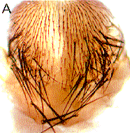
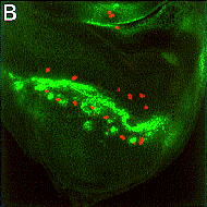
In Drosophila, decapentapledgic (dpp) encodes a homologue of BMP. We have been studying the involvement of dpp in the formation of mechanosensory sensory organ. Ectopic activation of Dpp signal by a constitutively activated form of a dpp receptor tkv Dpp receptor tkv increased the number of machanosensory organ. It was found that Dpp signal suppresses expression of another growth factor wingless. As a result, dpp induces sensory organ precursor (SOP) cells near by wingless expressing region. Position of SOP cells seems to be determined by the cooperation of at least two secreted growth factors, dpp and wingless.

Figure 2.
Interaction of dpp and wingless in the formation of the mechnosensory organ of Drosophila (a) increased mechanosensory bristles in the nortal region by ubiquitous expression of a constitutively active Dpp receptor Tkv. (b), induction of SOP cells (shown in red) nearby wingless expression domain (shown in green).


Publication List:
Akiyama, S., Katagiri, T., Namiki, M., Yamaji, N., Yamamoto, N., Miyama, K., Shibuya, H., Ueno, N., Wozney, J. M. , Suda, T. (1997) Constitutively active BMP type I receptors transduce BMP-2 signals without the ligand in C2C12 myoblasts. Exp. Cell. Res. 15, 362-369.
Higashijima, S. , Okamoto, H., Ueno, N., Hotta, Y. and Eguchi, G. (1997) High-frequency generation of transgenic zebrafish which reliably express GFP In whole muscles or the whole body by using promoters of zebrafish origin. Dev. Biol. 192, 289-299
Miya, T., Morita, K., Suzuki, A., Ueno, N. and Satoh, N. (1997) Functional analysis of an ascidian homologue of vertebrate Bmp-2/Bmp-4 suggests its role in the inhibition of neural fate specification. Development 124, 5149-5159.
Mochii, M., Ono, T., Matsubara, Y. and Eguchi, G. (1998) Spontaneous transdifferentiation of quail pigmented epithelial cell is accompanied by a mutation in the Mitf gene. Dev. Biol. 193, 47-62.
Namiki, M., Akiyama, S., Katagiri, T., Suzuki, A., Ueno, N., Yamaji, N., Rosen, V., Wozney, J. M., Suda, T. (1997) A kinase domain-truncated type I receptor blocks bonemorphogenetic protein-2-induced signal transduction in C2C12 myoblasts. J. Biol. Chem. 272, 22046-22052.
Natsume, T., Tomita, S., Iemura, S., Kintou. N., Yamaguchi, A. and Ueno, N. (1997) Interaction between soluble type I receptor for bone morphogenetic protein and bone morphogenetic protein-4. J. Biol. Chem. 272, 11535-11540.
Nikaido, M., Tada, M., Saji, T. and Ueno, N. (1997) Conservation of BMP signaling in zebrafish mesoderm patterning. Mech. Dev. 61, 75-88.
Shibuya, H., Iwata, H., Masuyama, N., Gotoh, Y., Yamaguchi, K., Irie, K., Matsumoto, K., Nishida, E. and Ueno, N. Role of TAK1 and TAB1 in BMP signaling in early Xenopus development. (1998) EMBO J. 17, 1019-1028.
Suzuki, A., Kaneko, E., Maeda, J., Ueno, N. (1997) Mesoderm induction by BMP-4 and -7 heterodimer. Biochem. Biophys. Res. Commun. 232, 153-156.
Suzuki, A., Kaneko, E., Ueno, N., Hemmati-Brivanlou, A. (1997) Regulation of epidermal induction by BMP2 and BMP7 signaling. Dev. Biol. 189, 112-122.
Suzuki, A., Ueno, N., and Hemmati-Brivanlou, A. (1997) Xenopus msx1 mediates epidermal induction and neural inhibition by BMP4. Development 124, 3037-3044.
Tonegawa, A., Funayama, N., Ueno, N., Takahashi, Y. (1997) Mesodermal subdivision along the mediolateral axis in chicken controlled by different concentrations of BMP-4. Development 124, 1975-1984.


webmaster@nibb.ac.jp
Last Modified: 12:00, May 28, 1998