NATIONAL INSITUTE FOR BASIC BIOLOGY  


National Institute for Basic Biology

Division of Molecular Neurobiology


Professor:
Masaharu Noda
Research Associates:
Nobuaki Maeda
Masahito Yamagata
Eiji Watanabe
NIBB Fellow:
Masakazu Takahashi
Graduate Students:
Hiroki Hamanaka (- Sept. 30, 1996)
Junichi Yuasa
Takafumi Shintani
Taeko Nishiwaki
Chika Saegusa (Oct. 1, 1996 -)
Visiting Scientists:
Ikuko Watakabe (Oct. 1, 1996 -)
Tatsunori Yamamoto* (- Aug. 31, 1996)
Technical Staffs:
Akiko Kawai
Shigemi Ohsugi
(*from Nagoya University)


We study the molecular and cellular mechanisms underlying the development of the vertebrate central nervous system. Our experiments are designed to analyze molecules and structures involved in regulation of various cellular events in brain morphogenesis such as generation of neuroblasts, their migration to form laminar structures and various nuclei, elongation and path-finding of neural processes, and the formation and refinement of specific connections between neurons. These studies utilize various techniques, including molecular biological (e.g. cDNA cloning, site-directed mutagenesis), biochemical (protein, carbohydrate) and immunological methods (monoclonal-antibody production), in addition to neuroanatomy, cell and organotypic culture (immortalized-cell production), and embryo manipulation (classical embryology, gene transfer with viral vectors, and gene targeting).


I. Retinotectal projection map and synapse formation

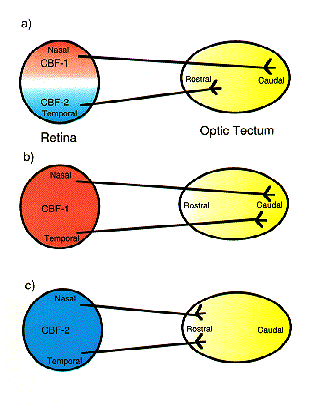
Topographic maps of neuronal connectivity have been reported for various parts of the nervous system. In the visual system of birds, retinal ganglion cell axons from the nasal (anterior) retina connect to a caudal (posterior) part of the midbrain visual center, the optic tectum, and temporal (posterior) retinal axons connect to the rostral (anterior) part, thereby establishing a point-to-point projection map. To understand the development of the retinotectal projection map, Sperry formulated the chemoaffinity theory in 1963, which is generally accepted today. His notion of chemoaffinity consists of five elements: (1) neurons are intrinsically different from each other, (2) these differences are position-dependent, (3) the differences are acquired very early, independently of their connection partners, (4) the differences are biochemical in nature, and (5) presynaptic and postsynaptic cells with matching biochemical labels connect with one another in a specific manner. Subscribing to this hypothesis, we employed a subtractive hybridization technique to identify molecules that display asymmetrical distributions between the nasal and the temporal retina. We discovered several distinct transcripts which are topographically expressed in the retina. Among these position-specific molecules, two winged-helix transcriptional regulators named CBF-1 and CBF-2 were expressed in the nasal and temporal retina, respectively. The winged-helix domain is a DNA-binding motif found in a family of transcription factors such as Drosophila forkhead and vertebrate HNF-3. By in situ hybridization, it was revealed that CBF-1 and CBF-2 transcripts began to be detected topographically in the primordial retina by embryonic day (E) 2, before birth of the retinal ganglion cells. Their topographic expression in the retina ceased by E10, prior to the onset of retinotectal connections. Misexpression of each factor in the retina using a replication-competent retroviral vector caused misprojection on the tectum along the rostro-caudal axis, suggesting that they determine the naso-temporal axis of the retina, and consequently specify the topographic projection of the retinal ganglion cell axons to the tectum by controlling expression of their target genes (Fig. 1). Our results provide evidence for the chemoaffinity theory, and this approach is promising for systematic discovery of a series of topographic molecules, determination of their functional hierarchy, and elucidation of the molecular and cellular mechanisms that generate the topographic map.

After reaching their appropriate sites along the rostro-caudal and dorso-ventral axes of the tectum, retinal axons begin to seek their appropriate laminar termination sites among 15 distinct laminae within the tectum. Our second aim is to identify molecular and cellular cues for ingrowing axons to arborize in appropriate laminae, to recognize particular postsynaptic partners, and finally to establish functional synaptic connections. Molecular and cellular studies for this purpose are currently underway.

Fig. 1.
CBF-1 and CBF-2 specify the retinotectal projection map
(a) CBF-1 and CBF-2 are expressed in the nasal and temporal retina, respectively. (b, c) Recombinant retrovirus-mediated misexpression of each factor changed the retinotectal projection map.


II. Receptor-like protein tyrosine phosphatases and brain development

Protein tyrosine phosphorylation is implicated in the various aspects of brain development. The pattern and level of tyrosine phosphorylation of the cellular proteins are controlled by a series of protein tyrosine kinases and protein tyrosine phosphatases (PTPs). In recent years, many classes of receptor-like PTPs (RPTPs) have been cloned and characterized. In 1994, we identified a brain-specific extracellular chondroitin sulfate proteoglycan, 6B4 proteoglycan/phosphacan as a splice variant of a proteoglycan-type RPTP, PTPz/RPTPb. PTPz and 6B4 proteoglycan have been suggested to play some roles in neurite outgrowth, neuronal cell migration and synapse formation. To understand the functions of these molecules, it is crucial to dissect the molecular bases of the signal transductions. Therefore, we attempted firstly to identify the ligands of PTPz.

Since 6B4 proteoglycan was considered to be identical to the extracellular portion of PTPz including carbohydrate modifications, we used this molecule for affinity chromatography to purify PTPz-binding proteins. From the CHAPS extract of rat brain microsomal fractions, 18-, 28-, and 40-kDa proteins were specifically isolated using 6B4 proteoglycan-Sepharose. N-terminal amino acid sequencing identified the 18-kDa protein as pleiotrophin/heparin-binding growth-associated molecule (HB-GAM), which was originally isolated from the brain as a mitogenic and neurite-promoting factor. Scatchard analysis of 6B4 proteoglycan-pleiotrophin binding revealed low (Kd = 3 nM) and high (Kd = 0.25 nM) affinity binding sites. Chondroitinase ABC digestion of the proteoglycan reduced the binding affinities to a single value (Kd = 13 nM) without affecting the number of binding sites. This suggested the presence of two subpopulations of the proteoglycan with different chondroitin sulfate structures. The binding of 6B4 proteoglycan to pleiotrophin was inhibited differently by various chondroitin sulfates. Interestingly, in contrast to chondroitin sulfate C which strongly inhibited binding of 6B4 proteoglycan to pleiotrophin (IC50 = 400 ng/ml), chondroitin sulfate A had almost no effect. These results suggested that chondroitin sulfate chains participate in the ligand-receptor interaction.

Immunofluorescence analysis indicated that both 6B4 proteoglycan and PTPz are localized on cortical neurons especially at the growth cones of their extending neurites. Anti-6B4 proteoglycan antibody added to the culture medium strongly suppressed pleiotrophin-induced neurite outgrowth of cortical neurons. Chondroitin sulfate C, but not chondroitin sulfate A, potently inhibited pleiotrophin-induced neurite outgrowth, consistently with observations regarding inhibition of the ligand-receptor interaction. These results suggested that pleiotrophin is a functional ligand for PTPz.

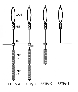
PTPz has another family member, RPTPg, which shows a high degree of structural similarity to PTPz. Pleiotrophin also has another family member, midkine, which also has mitogenic and neurite-outgrowth promoting activities. Therefore, the ligand-receptor relationship between pleiotrophin, midkine and PTPz, RPTPg is an important question which should be addressed. As the first step, we cloned RPTPg cDNAs to reveal its molecular diversity. cDNA clones for the four splicing variants of RPTPg were isolated from rat brain cDNA libraries (Fig. 2). We designated these molecules as RPTPg-A, -B, -C and -S. RPTPg-A was the longest form and had a similar structure to human and mouse RPTPg. RPTPg-B was devoid of the intracellular juxtamembrane 29 amino acids of RPTPg-A. Recently, this type was independently found in a human kidney-derived cell line, ACHN, by Sorio et al. The other two variants were novel: RPTPg-C had a single phosphatase domain, and RPTPg-S was a secretory type of RPTPg. mRNAs of these four species were expressed in the brain, kidney, lung and heart. Transfection of RPTPg-A and -S expression plasmids into COS7 cells resulted in the expression of membrane-bound 190- and secretory 120-kDa proteins, respectively. RPTPg is thus comparable to PTPz with regard not only to structure but also to the presence of both secretory and transmembrane forms. However, any RPTPg variants ectopically expressed in these cells did not exist as proteoglycans contrasting to PTPz variants.

Fig. 2.
Schematic representation of RPTPg isoforms
RPTPg consists of a carbonic anhydrase-like domain (CAH; open circles), a fibronectin type III-like domain (FN III; hatched boxes), a transmembrane segment (TM; open boxes) and two tyrosine phosphatase domains (PTP-DI and -DII; shaded boxes). The dashed line indicates the deleted sequence in RPTPg-B. RPTPg-C possesses only a single tyrosine phosphatase domain, and RPTPg-S is an extracellular variant of RPTPg.


III. Gene targeting of Na-G, a voltage-gated sodium channel expressed in glial cells

Glial cells have been considered to be inexcitable. Despite the lack of electrical excitability, they express voltage-gated sodium channels with properties similar to the sodium channels in excitable cells. The cellular function of these voltage-gated sodium channels is not clear. An in vitro study raised the possibility that glial sodium channels serve as a pathway for sodium-ion entry to fuel Na-K ATPase, which requires three sodium ions for transport of two potassium ions. A drop in the intracellular concentration of sodium ion concentration through these channels may hamper ATPase activity. To test this hypothesis and clarify the roles of glial sodium channels in vivo, we are currently attempting to generate knock-out mice deficient in the glial voltage-gated sodium channel, Na-G, the cDNA of which was recently cloned. We have successfully produced chimeric mice which contain the targeted embryonic stem-cells contribution.

By applying gene targeting technology to other novel genes expressed in the brain, we intend to investigate the molecular mechanisms underlying the development and function of the brain.


Publication List:
1. Original Papers
Leber, S. M., Yamagata, M. and Sanes, J. R. (1996) Gene transfer using replication-defective retroviral and adenoviral vectors. In Methods in Avian Embryology (Bronner-Fraser, M. ed., Methods in Cell Biology, Academic Press, Orlando) 51, 161-183.
Maeda, N. and Noda, M. (1996) 6B4 proteoglycan/ phosphacan is a repulsive substratum but promotes morphological differentiation of cortical neurons. Development 122, 647-658.
Nishizuka, M., Ikeda, S., Arai, Y., Maeda, N. and Noda, M. (1996) Cell surface-associated extracellular distribution of a neural proteoglycan, 6B4 proteoglycan/phosphacan, in the olfactory epithelium, olfactory nerve, and cells migrating along the olfactory nerve in chick embryos. Neurosci. Res. 24, 345-355.
Watanabe, E., Matsui, F., Keino, H., Ono, K., Kushima, Y., Noda, M. and Oohira, A. (1996) A membrane-bound heparan sulfate proteoglycan that is transiently expressed on growing axons in the rat brain. J. Neurosci. Res. 44, 84-96.
Watanabe, E., Kushima, Y. and Oohira, A. (1996) Heparan sulfate proteoglycan associated with growing parallel fibers of granule cells in the rat cerebellar cortex. J. Brain Sci. 22, 67-75.
Maeda, N., Nishiwaki, T., Shintani, T., Hamanaka, H. and Noda, M. (1996) 6B4 proteoglycan/phosphacan, an extracellular variant of receptor-like protein tyrosine phosphatase z/RPTPb, binds pleiotrophin/HB-GAM. J. Biol. Chem. 271, 21446-21452.
Yuasa, J., Hirano, S., Yamagata, M. and Noda, M. (1996) Visual projection map specified by expression of transcription factors in the retina. Nature 382, 632-635.
Shintani, T., Maeda, N., Nishiwaki, T. and Noda, M. (1997) Characterization of rat receptor-like protein tyrosine phosphatase g isoforms. Biochem. Biophys. Res. Comm., 230, 419-425.
2. Proceedings
Maeda, N. and Noda, M. (1996) 6B4 proteoglycan/phosphacan and development of the brain. In "Generation of Neuronal Diversity and Specificity in the Brain", pp. 66-70. (Taniguchi International Symposium)
Yamagata, M. and Noda, M. (1996) Layer-specific neuronal connections in the retinotectal system. In "Generation of Neuronal Diversity and Specificity in the Brain", pp. 101-103. (Taniguchi International Symposium)
Yamagata, M., Takahashi, M. and Noda, M. (1996) Expression of ezrin in a subset of the chick retinotectal projection. (Society for Neuroscience Abstract)


nibb-adm@nibb.ac.jp
Last Modified: 12:00, June 27, 1997