NATIONAL INSITUTE FOR BASIC BIOLOGY
![]()
National Institute for Basic Biology
1) from Nagoya University |
2) from Hiroshima University |
3) from Colombia National University |
![]() |
Fig. 1 Targeting and processing of the chimeric proteins between N-terminal sequence of glyoxysomal citrate synthase (gCS) and b-glucuronidase (GUS). (A) Localization of the chimeric proteins in transgenic Arabidopsis plants analyzed by immunoelectron microscopy. 13-day-old cotyledons were fixed and embedded in LR-white resin. The thin sections were stained with anti-GUS antibodies. Gold particles indicated the localization of the GUS chimeric proteins in microbodies. Bar in panel DC42 indicates 1 mm. (B) Construction of the chimeric proteins. The yellow and green boxes show the consensus sequences which were found in the N-terminal region of the microbody proteins that contained the cleavable N-terminal presequences. Mutated amino acids are indicated by red letters. (C) Summary of the localization and processing of the chimeric proteins. Arg-16, Leu-17 and Leu-24 in gCS, which are located in the yellow box, function in the targeting to microbodies, whereas Cys-42 located in the green box functions in the processing of the presequence. |
![]() |
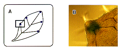
Fig. 2. Histochemical localization of GUS activity in the hydathode of Arabidopsis transformed with the g-VPE promoter-gus fusion gene. Arabidopsis was transformed with a reporter gene composed of the promoter of the g-VPE gene and the coding region of b-glucuronidase (GUS). GUS activity was observed in the hydathodes at the ends of veins along with the leaf margins, as shown in (A). Hydathodes are structures for that discharge water from the interior of the leaf to its surface. A photograph of the hydathode with DIC optic of the enclosed region in A is shown in (B). |
nibb-adm@nibb.ac.jp
Last Modified: 12:00, June 27, 1997