National Insitute for Basic Biology  


DIVISION OF MORPHOGENESIS


Professor:
Goro Eguchi
Associate Professor:
Ryuji Kodama
Research Associates:
Makoto Mochii
Mitsuko Kosaka
Visiting Scientists:
Takamasa S. Yamamoto
Kaichiro Sawada
Akio Iio
Toshiyuki Nagamoto
Graduate Students:
Nobuhiko Mizuno (Graduate University for Advanced Studies)
Yuuichi Mazaki (Graduate University for Advanced Studies)
Harutoshi Hayashi (from School of Agriculture, University of Tokyo)
Jatupol Kositsawat (from School of Medicine, University of Tokyo)
Technical Staffs:
Chiyo Takagi
Hisae Urai
In the newt and some other limited animal species, the lens and neural retina can be completely regenerated through transdifferentiation of pigmented epithelial cells (PECs). Such a phenomenon, transdifferentiation, as observed in regeneration of ocular tissues seems to be a highly powerful model for studying stability and instability in differentiation of tissue cells. From this view point, lens transdifferentiation of PECs of vertebrate has been studied in vivo and in vitro systems, and our in vitro studies have revealed that dormant potential to transdifferentiate into lens cells is widely conserved throughout vertebrate species, and that the cell type-specific genes are completely inactivated in the multipotent (at least bipotent) dedifferentiated cells originated from pigmented epithelial cells.

Our studies have been conducted to clarify the molecular mechanism controlling the lens transdifferentiation in vertebrate PECs and also to search the reason why the pigmented epithelia of species other than the newt and so forth never regenerate the lens in the in situ eyes. Based on findings accumulated up to the last year, we have conducted analysis of the lens transdifferentiation of PECs in vivo and in vitro and the following results have been established.


I. Multipotency conserved in the human PECs

It has been proved that retinal PECs isolated from the eye of 80-year-old human can transdifferentiate into lens cells to construct lentoids when dissociated and cultured under the culture condition established in our laboratory (cf. Itoh and Eguchi, Dev. Biol. 115: 353-362, 1986). Human PECs can readily transdifferentiate and form lentoids, which express major human crystallin genes and ultrastructures characteristic to lens, through the dedifferentiated state in the similar manner as observed in chicken retinal PECs. We have isolated clones from the progeny of dedifferentiated human retinal PECs to establish cell lines. Reaggregates of cells of one of established cell lines were found to express lens specificities when maintained under the culture condition permissive for lens cell differentiation (cf. Itoh and Eguchi, 1986), although these dedifferentiated cells could not develop to lentoid in the monolayer culture. However, when maintained with the ordinary medium, Eagle's minimum essential medium supplemented with 10% of fetal bovine serum, for long time, they express neuronal cell morphology (Fig. 1). Neuronal specificities of these cells with neuronal morphology could be detected by indirect immunofluorescent staining using antibodies against 70, 150 and 200 kd subunits of neurofilament protein. This result is the first evidence that human retinal PECs can express neuronal phenotype by themselves and is strongly suggesting that the human retinal PECs, like the newt PECs, conserve multipotency for differentiation, although neuronal transdifferentiation of human retinal PECs by transfection with H-ras proto-oncogene has been reported by Dutt, Scott et al (DNA and Cell Biology 12(8): 667-673, 1993). The human PECs grow well and endure genetic manipulations, such as gene transfection, thus are supposed to be a promising material to study transdifferentiation. We are now extending our studies of transdifferentiation using this cell line established from human retinal PECs.

Fig.1

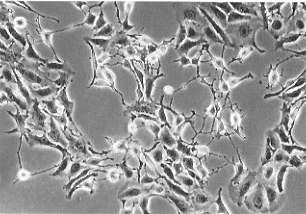
Fig.1
Human retinal PECs showing a neuron-like morphology. Cells were isolated from the eye of an 80-year-old donor and their growth was enhanced by the EdFPH medium. Cells were then cultured in the standard medium for about30 days.


II. Establishment of in vitro model system of lens transdifferentiation using chicken iris PECs

As the main material to study the process of transdifferentiation in our laboratory, we have been utilized retinal PECs isolated from approximately 10 day-old chick embryos, because we can obtain large number of PECs through an easy manipulation, owing to the large size of the eye of the chick embryo. Recently, however, we have come to recognize that cell culture of the retinal PECs has gradually become difficult. Cells are susceptible to the change of culture conditions, such as the product lot of the fetal calf serum or other components of the culture medium, and often stop growing, embedded in irregularly accumulated extracellular matrices. Besides trials to modify culture conditions to enable more efficient culture of the retinal PECs, other sources of PECs were also sought. The iris PECs of the chick has emerged as a promising material as briefly described below.

Because it is the iris PECs that give rise to lens regeneration in the newt, the iris PECs of the chicken was tested for their ability to transdifferentiate in vitro. The iris was isolated from newly hatched chicks, and the pigmented epithelium was cleanly separated from the stroma using dispase. The iris PECs slowly grow and stably maintain the phenotype in a standard culture medium. When the EdFPH medium, which is effective in inducing dedifferentiation of retinal PECs (cf. Itoh and Eguchi, 1986), was applied, the iris PECs grow rapidly and dedifferentiate. By further addition of ascorbic acid, such dedifferentiated cells accumulate and form large number of lentoids (Fig. 2). The growth and transdifferentiation of the iris PECs are highly reproducible. Preliminary studies have shown that all the genes we have analyzed in the retinal PECs are expressed in the culture of the iris PECs.

Fig.2

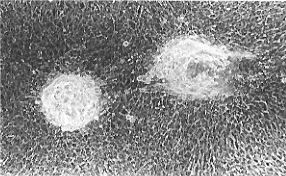
Fig.2
Lentoids formed in the culture of the iris PECs of newly hatched chicks. Cell passaged in the EdFPH medium for several times were cultured with ascorbic acid for about 30 days.


III. Detailed analysis of the gap junctions in the Wolffian lens regeneration

On the basis of the results accumu-lated through the studies of lens transdifferentiation of the retinal PECs of the chick embryo, detailed studies of the mechanisms regulating the lens regeneration in the newt have become possible. Our recent observation in the chick PECs has shown that the dePECs are devoid of both the structure and conductance of the gap junction. There was also a preliminary study that showed a transient absence of the electric coupling between dorsal iris PECs prior to the lens regeneration. We have started a study on the gap junction between the iris PECs during lens regeneration, and have obtained preliminary results shown below.

A significant decrease in the number of gap junctions was detected in the dorsal iris pigmented epithelium, but not in the ventral, on approximately 10 days after lens removal. This result confirms the preliminary result in the electric coupling, and is in accordance with the result in the chick retinal PECs. However, we also found that, just after the lens removal, there is a period during which gap junctions are increased all over the iris. This increase was accompanied with the increase in the transcript of the connexin gene, which codes for a major component of the gap junction (R. Kodama, unpublished).

Similar study in the partial hepatectomy showed that, shortly after the hepatectomy, there is a quick decrease in gap junction number, which is then followed by the raise in DNA synthesis and mitoses. Our result in the lens regeneration suggests that the regeneration process is biphasic, consisting of a period of emergency repair in the whole iris and a period of regeneration only in the dorsal iris. Through such renewed studies, which are also focused on growth factors and tissue proteases, a clearer image of the lens regeneration process will be obtained.


Publication List:

Kodama, R. and Eguchi, G. (1994) The loss of gap junctional cell-to-cell communication is coupled with dedifferentiation of retinal pigmented epithelial cells in the course of transdifferentiation into the lens. Int. J Dev. Biol. 38, 357-364.

Kodama, R. and Eguchi, G. (1994) Gene regulation and differentiation in vertebrate ocular tissues. Curr. Opin. Genetics Dev. 4, 703-308.

Iio, A., Mochii, M., Agata, K., Kodama, R. and Eguchi, G. (1994) Expression of the retinal pigmented epithelial cell-specific pP344 gene during development of the chicken eye and identification of its product. Develop. Growth Differ. 36, 155-164.

Ono, T., Murakami, T., Mochii, M., Agata, K., Kino, K., Otsuka, K., Ohta, M., Mizutani, M., Yoshida, M. and Eguchi, G. (1994) A complete culture system for avian transgenesis, supporting quail embryos from the single-cell stage to hatching. Dev. Biol. 161, 126-130.

Ono, T., Muto, S., Mizutani, M., Agata, K., Mochii, M., Kino, K., Otsuka, K., Ohta, M., Yoshida, M. and Eguchi, G. (1994) Production of quail chimera by transfer of early blastodermal cells and its use for transgenesis. Jap. Poultry Sci. 31, 119-129.

Orii, H., Hyuga, M., Mochii, M., Kosaka, J., Eguchi, G. and Watanabe, K. (1994) Predominant melanogenesis and lentoidogenesis in vitro from multipotent pineal cells by dimethyl sulfoxide and hexamethylene bisacetamide. Int. J. Dev. Biol. 38, 397-404.