(1)from Tokyo University)
(2)from Aichi University of Education)
Brain can be seen as an integrated circuit where neurons of various identities are interconnected in a highly ordered manner by their axons. We have been interested in how individual neurons aquire their own identities and how their axons find their own pathways and finally recognize their proper targets. Using two different animals, zebrafish (Danio rerio) and fly (Drosophila melanogaster), both of which are suitable for genetic analysis and gene manipulation, we are trying to address these questions both at the molecular and cellular levels.
I. Differential expression of islet-1 homologs during specification of primary motoneuron
Islet-1 (Isl-1) is a LIM domain/homeodomain-type transcription regulator originally identified as an insulin gene enhancer binding protein. Isl-1 is considered to be involved in the differentiation of the neuronal cells. We previously cloned a isl-1 homolog from zebrafish cDNA Iibrary, named it zebrafishisl-1. Recently, we have isolated two novel isl-1 homologs from zebrafish cDNA Iibrary, named them zebrafishisl-2 and 3.
We examined the mRNA expression pattern of each homolog using in situ hybridization to whole-mount embryos. All three homologs are expressed in Rohon-Beard neurons. However the expression
in primary motoneurons diverged. Zebrafishisl-1 mRNA is expressed in the rostral primary motoneuron (RoP). isl-2 mRNA is expressed in the caudal primary motoneuron (CaP) and its variant sibling (VaP). isl-3 mRNA is expressed in the ventral region of the myotome but not in the primary motoneurons. The ventral myotome is the region that the axon of the CaP extend into. isl-3 mRNA is also expressed throughout the developing eye and tectal region of the midbrain, the target for the retial axons. These results raise possibilities that the isl-1 homologs may be involved in the specification and/or target recognition by the primary motoneurons.
II. Cloning and expression of AN34/F-Spondin family in zebrafish
The floor plate is a cell group implicated in the control of neural cell pattern and axonal growth in the developing vertebrate CNS. By subtractive hybridization technique, Klar et al. identified F-Spondin, a novel secreted protein ex-pressed at high levels in the floor plate. The C-terminal half of the protein contains six TSRs (Thrombospondin Type 1 repeats), while the N-terminal half exhibited no homology to other proteins. Although F-Spondin was shown to promote neural cell adhesion and neurite extension in vitro, its functions in vivo remain largely unknown.
Nose performed an enhancer trap screen to search for genes expressed by a subset of muscle cells in Drosophila. One of such genes, AN34 was cloned and found to encode a secreted protein sharing homology in two parts (H1 and H2) with the N-terminal half of F-Spondin (Nose, personal communication).
We expected the homologous reigions to be novel domains important for neuronal development, and thus, searched for AN34/F-Spondin family from zebrafish (Danio rerio). We identified two novel genes (zfAN1 and zfAN2) in addition to zf-F-Spondin. zfAN1 and zfAN2 consist of H1, H2 and one TSR. The overall structure are highly similar to AN34 and more related to AN34 than F-Spondin, suggesing that zfANl, zfAN2 and AN34 constitute a novel subfamily. zfAN1 is expressed weakly in the floor plate and a small subset of neuronal cells at 24-30 hr. zfAN2 expression is first observed broadly around the axial mesoderm (notochord) at 10-12 hr. Then, the expression becomes restricted to the floor plate and hypochord at 14 hr and continues at high levels up to 30 hr. F-Spondin is expressed in the floor plate as seen in rat. These findings suggest that the AN34/F-Spondin family play important roles in the midline development in the vertebrate.
III. Search for new cell surface recognition molecules expressed in a subset of CNS axons
During neuronal development, growih cones are known to have ability to recognize and extend along specific axonal surfaces. Several lines of evidence suggest that cell surface glycoproteins play important roles in this process. Immunocytochemical studies have demonstrated that the monoclonal antibody HNK-1, originaly raised to a human lymphoblastoma, recognizes a subset of CNS and PNS axons in many vertebrates including zebrafish by binding to a carbohydrate determinant in several glycoproteins. Thus, we started to characterize glycoproteins recognized by HNK-1, hoping to isolate new cell adhesion molecules (CAMs) expressed in a subset of CNS and PNS axons during neuronal development.
Our strategy is as follows: Many brains of adult zebrafish are homogenized, and from this extract, molecules which binds to HNK-1 are intensely enriched by using HNK-1 affinity chromatography. The affinty-purified fractions are used to immunize mice to get a series of monoclonal antibodies (MAbs). The MAbs, thus obtained, are screened with immunohistochemistry to zebrafish tissues. Hopefully, those MAbs which recognize peptides and not carbohydrates are used to isolate and characterize new molecules.
Immunogen which represents several protein families on Western Blotting was prepared. Mice were immunized with it, and hybrydoma cells were generated by cell fusion. A few MAbs each of which recognizes different protein species have been obtained. One of them could recognize a subset of CNS axons in the adult brain. Efforts are currently underway to obtain more MAbs.
IV. Tol1: Tc1-like element of Oryzias latipes
The zebrafish and the medaka have the potential to become important organisms for molecular genetic analysis of early vertebrate development. This is because both fishes offer the possibility of combining genetics with excellent embryology. The goal of this research is to use transposon to perform mutant screens in these fishes in order to identify, and subsequently clone genes important in early development, especially in neurogenesis.
A transposon, Tc1, which moves via DNA intermediate has been a useful tool in C.elegans genetics. Recently homologs of Tc1 were found in some fishes, raising the possibility that insertional mutagenesis would also be possible in vertebrate.
We discovered a Tc1-like element in the genome of medaka. Southern hybridization analysis showed that the element, Tol1, exists as multi copy in medaka genome but did not exist in zebrafish genome. Although the element seems to encode a protein similar to the Tc1 family of transposes, the putative transposase is probably not functional due to mutations as other putative transposases encoded by Tc1-like elements in fishes.
V. Fasciclin III as a synaptic recognition molecule in Drosophila
The larval neuromuscular system of the Drosophila consists of uniquely identified cells, and is a powerful model system for studying selective synapse formation. During synaptogenesis, the cell adhesion molecule Fasciclin III appears in both motoneuron RP3 and its targets, muscle 6 and 7. We have tested whether fasciclin III is necessary and/or sufficient for RP3 targe selection using intracellular dye injection.
First, we have found that in the existing fasciclin III null mutant RP3 reliably formed synapse with its normal targets. Therefore, fasciclin III is either irrelevant for the process, or playing a positive role but its absence can be compensated for another redundant mechanism ("X").
We now have demonstrated that the latter is the case. We geherated transgenic flies which misexpresses fasciclin III extopically on all skeltal muscles during neuromuscular synaptogenesis. This was accompanied by creating a construct which placed the fasciclin III gene under the control of the myosin heavy chain promoter, and introducing this construct into the fly genome by P-element mediated genomic transformation. In these flies, RP3 often innervated non-target
muscle cells while other identified motoneurons innervated targets normally.
Our results provide the single identified-cell level evidence that a cell adhesion molecule functions as a specific synaptic target recognition molecule in vivo.
VI. no optic lobes (nol), a gene that is essential for larval neurogenesis in Drosophila
We have identified a larval/pupal lethal line by P-element insertion mutagenesis. Although embryonic neurogenesis of the homozygous mutant is fairly normal, BrdU incorporation analysis revealed they lack proliferating larval neuroblasts and some non-neuroblasts cells (neurons?) start DNA synthesis at late larval stage. On the other hand, imaginal discs differentiate normally, so that rare escapers become normal looking pupae with a small central nervous system. The most characteristic feature is that they have no optic lobes. In such an individual, retinular axons lose their targets and are directly guided into the tiny brain. We conclude that there is an interesting gene named as no optic lobes (nol) near the P-insertion, which is essential for proliferation and differentiation of larval neuroblasts.
We cloned genomic regions around the P-element to find a transcription unit whose expression is significantly reduced in the homozygotes. The putative nol gene encodes a novel protein with a hydrophobic signal sequence. The deduced amino acid sequence has a mucin-like region, but its significance remains to be analyzed.
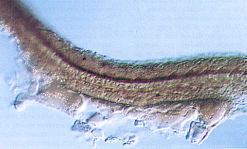
The expression pattern of F-Spondin mRNA in the spinal cord. F-Spondin mRNA is expressed in the floor plate cells.
Publication List:
Inoue, A., Hatta, K., Hotta, Y. and Okamoto, H. (1994) Developmental regulation of Islet-1 mRNA expression during neuronal differentiation in embryonic zebrafish. Devel. Dynam.199, 1-11.
Goto, S., Tanimura, T. and Hotta, Y. (1995) Emhancer trap detection of positional information corresponding to the polar coordinate system in the imainal disc of Drosophila melanogaster. Roux's Archiv Develop. Biol. (In the Press).
Chiba, A., Snow, P., Keshishian, H. and Hotta, Y. (1995) Fasciclin III as a synaptic target recognition molecule in Drosophila. Nature374, 166-168.
National Insitute for Basic Biology