
|
|
DIVISION OF DEVELOPMENTAL GENETICS |
| Professor: | KOBAYASHI, Satoru |
| Research Professors: | MUKAI, Masanori |
| SHIGENOBU, Shuji | |
| Technical Staff: | NODA, Chiyo |
| Postdoctoral fellows: | SATO, Kimihiro |
| HAYASHI, Yoshiki | |
| Graduate Students: | KITADATE, Yu |
| HAYASHI, Makoto | |
| YATSU, Jun | |
| Technical Assistants: | ARITA, Kayo |
| UEDA, Yoshiko | |
| TSUCHIYA, Naomi | |
| SATO, Kaori | |
| Secretary: | HONDA, Satoko |
|
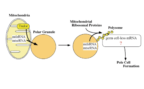
The germ cells are the specialized cells, which can transmit the genetic materials from one generation to the next in sexual reproduction. All the other cells of the body are somatic cells. This separation of germ and somatic cells is one of the oldest problems in developmental biology. In many animal groups, a specialized portion of egg cytoplasm, or germ plasm, is inherited by the cell lineage which gives rise to germ cells. This cell lineage is called germline. The germline progenitors eventually migrate into the gonads, where they differentiate as germ cells when the organisms are physically matured. Earlier investigators have demonstrated that germ plasm contains maternal factors required and sufficient for germline formation. In Drosophila, this cytoplasm is histologically marked by the presence of polar granules, which act as a repository for the maternal factor required for germline formation. Our molecular screens have identified several factors stored in the polar granules. One of the factors is mitochondrial large rRNA which functions to form the germline progenitors, or pole cells. The other is nanos mRNA, which is required for pole cell differentiation. I. Role of Mitochondrial Ribosomal RNAs in Pole Cell FormationUltrastructural studies have shown that the germ plasm is basically composed of polar granules and mitochondria. While the primary roles of the mitochondria are oxidative phosphorylation and biosynthesis of many metabolites, it has now become evident that they are also involved in germline formation. In Drosophila, pole cell formation requires the function of mitochondrial ribosomal RNA in germ plasm. We have previously reported that mitochondrial large rRNA (mtlrRNA) and small rRNA (mtsrRNA) are both transported from mitochondria to polar granules. This transportation occurs during early embryogenesis, when mitochondria are tightly associated with polar granules in germ plasm, and it depends on the function of the maternally-acting gene, tudor, that is known to be required for pole cell formation. Mitochondrial rRNAs remain on the polar granules until pole cell formation and are no longer discernible on the granules within pole cells. Reduction of the extra-mitochondrial mtlrRNA amount results in the failure to form pole cells and injection of mtlrRNA is able to induce pole cells in embryos whose ability to form these cells has been abolished by uv-irradiation. These observations clearly show that the extra-mitochondrial mtlrRNA on polar granules has an essential role in pole cell formation, presumably cooperating with mtsrRNA. |
 |
|
Fig. 1 Role of mitochondrial rRNAs in pole cell formation |
 |
|
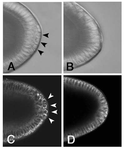
Fig. 2 Injection of kasugamycin causes a defect in pole cell formation but not in mitochondrial activity. Embryos at stage 4-5 were injected with DW (A, C) and KA (B, D) and photographed using a compound microscope equipped with Nomarski optics. (A, B) Pole cells are not discernible in the KA-injected embryo, although somatic cells are intact. (C, D) Injected embryos were stained with Rhodamine 123, which was not affected by KA treatment. We found that CH treatment also inhibited pole cell formation, but did not affect Rhodamine 123 staining (data not shown). Arrowheads in (A, C) indicate the pole cells. |
|
We have found that mitochondrial rRNAs form mitochondrial-type of ribosomes on polar granules, cooperating with mitochondrial ribosomal proteins. This suggests the possibility that the protein(s) essential for pole cell formation is produced by the mitochondrial-type of ribosomes (Fig. 1). To address this issue, we have been examining the effect of Chloramphenicol and Kasugamycin (KA) on pole cell formation. Chloramphenicol and Kasugamycin are known to inhibit mitochondrial (prokaryotic)-type of translation. When these antibiotics were injected into the posterior pole region of early embryos, pole cell formation was severely affected (Fig. 2). In contrast, Chloramphenicol and Kasugamycin treatment did not affect somatic cell formation at a dose we used. These observations strongly suggest that the mitochondrial-type of translation system must be intact for the embryos to form pole cells. Recently, we found that one of the target RNAs translated on the mitochondrial-type of ribosomes was germ cell-less (gcl) mRNA, which is stored in polar granules and encodes a protein required for pole cell formation. |
 |
|
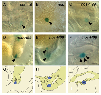
Fig. 3 nos-H99 pole cells have the ability to adopt somatic cell fate and to enter into the embryonic gonads. Pole cells from nos (B), nos-H99 (C-F) and control embryos (A) were transplanted into hosts, and the hosts were developed until stage 15-17. The transplanted pole cells were identified as blue cells after heat treatment and staining with X-gal (arrowheads). The transplanted control (A) and nos-H99 pole cells (C) were observed within the gonads, while nos pole cells remained outside the gonads (B). Some nos-H99 pole cells were integrated within somatic tissues (D-F). (D) nos-H99 pole cell within midgut epithelium is shown. Note that nos-H99 pole cell and the neighboring midgut epithelial cells were columnar in shape at stage 17. nos-H99 pole cells within gastric caeca (E) and tracheal epithelium (F) are also shown. The lower cell pointed by arrowhead in (E) was out of focus. G-I, Drawings tracing the integrated nos-H99 pole cells (blue) and the surrounding somatic cells in (D-F), respectively. Scale bar; 10 µm. |
II. Role of Nanos protein in pole cell differentiationPole cells differ from the soma in regulation of transcriptional activity. Pole cells are transcriptionally quiescent until the onset of gastrulation, although transcription is initiated in the soma during the syncytial blastoderm stage. Consistent with this, RNA polymerase II (RNAP II) remains inactive in early pole cells. Thus, the ability to express zygotic mRNA-encoding genes is suppressed only in pole cells in early embryos. |
|
|
|
|
 |
|
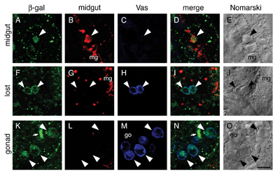
Fig. 4 nos-H99 pole cells within midgut epithelium expressed midgut marker genes but lost germline marker, Vas. We examined expression of midgut marker genes and Vas protein in nos-H99 pole cells within midgut epithelium (A-E), haemocoel (“losta ” pole cells) (F-J) and the gonad (K-O) of host embryos. The transplanted pole cells were identified as the cells expressing β-gal (marked by arrowheads). The embryos at stage 13-15 were triple-stained with probes for the midgut marker genes (bInt-n/mex1;red) (B, G, L) and antibodies against β-gal (green) (A, F, K) and Vas protein (blue) (C, H, M). nos-H99 pole cell integrated within midgut epithelium (mg), like the neighboring host cells, was cuboidal in shape at stage 14 (E). The a &idquo;integrated” pole cell expressed midgut marker genes (B), but not Vas protein (C). In contrast, nos-H99 pole cells, which were found within haemocoel in the vicinity of midgut (mg) (J), maintained Vas expression (H), but never expressed midgut marker genes (G). Within the gonad, nos-H99 pole cells and the host pole cells were both negative for midgut markers (L). Arrows in (K) and (N) indicate non-specific staining of tracheal lumen. Merged images (D, I, N) and photographs taken with a compound microscope equipped with Nomarski optics (E, J, O) are shown. Dotted lines indicate midgut epithelia (mg) in (E) and (J), and the gonad (go) in (O). Scale bar; 10 µm. |
|
Among the maternal components of germ plasm, Nanos (Nos) is essential for the germline-specific events occurring in pole cells. nos mRNA is localized in the germ plasm during oogenesis, and is translated in situ to produce Nos protein after fertilization. Nos is only transiently present in the posterior half of embryos during the preblastoderm stage, and is required there for posterior somatic patterning. Nos in the germ plasm is more stably inherited into the pole cells at the blastoderm stage, remaining detectable in these cells throughout embryogenesis. Pole cells that lack Nos (nos- pole cells) are unable to follow normal germline development; they fail to migrate properly into the embryonic gonads, and consequently do not become functional germ cells. nos- pole cells fail to establish and/or maintain transcriptional quiescence, and ectopically express somatically-transcribed genes, including fushi tarazu(ftz), even-skipped (eve) and Sex-lethal (Sxl). Nos represses translation of mRNAs with discrete RNA sequences called Nos response elements (NREs). In the pathway leading to posterior somatic patterning, Nos acts together with unlocalized Pumilio (Pum) protein to repress translation of maternal hunchback (hb) mRNA. This translational repression is mediated by binding of Pum to NREs in the 3'-untranslated region (UTR) of hb mRNA. In pole cells, Nos also acts with Pum to regulate germline-specific events. Pum, like Nos, is required in pole cells for their migration to the gonads. We have found that Nos, along with Pum, represses translation of importin α2 (imp α2) mRNA in early pole cells. The impα2 mRNA contains an NRE-like sequence in its 3'-UTR and encodes a protein required for nuclear import of karyophilic proteins. We found that Nos inhibits expression of a somatically-transcribed gene, ftz, in pole cells by repressing Impα2-dependent nuclear import of a transcriptional activator for ftz, Ftz-F1. The above results raise the question whether the pole cells lacking Nos(nos- pole cells) are able to differentiate into somatic cells. However, it is difficult to study their developmental fate, since Nos also represses apoptosis of pole cells, and almost all of nos- pole cells are eliminated until at least the end of embryogenesis. To overcome this problem, we used Df(3L)H99, a deletion for three genes required for apoptosis. Introduction of the H99 deficiency results in nos- pole cells being escaped from apoptosis. We transplanted the nos--H99- pole cells into normal embryos and observed their behavior, and found that some of nos--H99- pole cells were able to differentiate as somatic cells (Fig. 3, 4). This suggests that pole cells have the ability to differentiate as somatic cells, but its ability is inhibited by Nanos activity. Recently, we have found that somatic differentiation of nos--H99- pole cells requires Impα-2 activity, suggesting that Nos inhibits somatic differentiation by repressing Impα-2 production. III. Comprehensive analysis of genes expressed in Drosophila gonad |
|
|
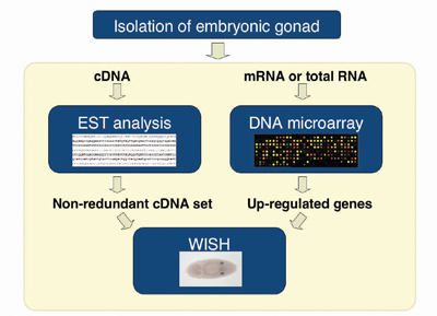
Fig. 5 Transcriptome analysis of the embryonic gonads |
|
|
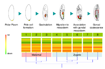
Fig. 6 Clustering analysis of expression profile of all of the predicted genes in the Drosophila genome. We divided embryonic stages into 8 groups, and isolated pole cells from the embryos at each stage. These pole cells were used for microarray analysis. As a reference, we used RNA from whole embryos of these 8 groups. Single line represents single transcript. Red line shows a “upregulateda” in pole cells. Hierarchical clustering analysis shows that they can be divided into two large groups. This is due to the onset of zygotic transcription in group-4 pole cells. |
|
To explore the regulatory mechanism of germline specification, we attempted to identify genes expressed in pole cells and/or in somatic cells within the gonad by a comprehensive approach. From the embryos carrying EGFP-vasa transgene that express GFP only in pole cells, we isolated the gonads by using fluorescence-activated cell sorting (FACS), and costructed a gonad cDNA library. Each cDNA clone was sequenced from both 5’ and 3’ ends, and these Expression Sequence Tags (ESTs) were computationally condensed into sequence clusters, which were then subjected to whole-mount in situ hybridization (WISH) (Fig. 5). Approximately 20,227 of ESTs were generated, and were clustered into 2900 distinct genes. The WISH analysis identified more than 130 genes that were expressed predominantly in the gonads. We also started to identify genes expressed predominantly in pole cells, using DNA microarray technique (Fig.6). |
Publication List: |
|
Hayashi, Y., Hayashi, M. and Kobayashi, S. (2004) Nanos suppresses somatic cell fate in Drosophila germline. Proc. Natl. Acad. Sci. USA. 101, 10338-10342. |
|
Hanyu-Nakamura, K., Kobayashi, S. and Nakamura, A. (2004) Intrinsic and extrinsic lipid phosphate phosphatase defines cell viability that promotes directional migration of Drosophila germ cells. Development 131, 4545-4553. |
|
Unezaki, S., Nishizawa, M., Okuda-Ashitaka, E., Masu, Y., Mukai, M., Kobayashi, S., Sawamoto, K., Okano, H. and Ito, S. (2004) Characterization of the isoforms of MOVO zinc finger protein, a mouse homologue of Drosophila ovom as transcription factors. Gene 336, 47-58. |
 |