RESEARCH SUPPORT FACILITY
 
Head of Facility:


Associate Professor:
Research Associates:

Technical Staffs:
MURATA, Norio (~Mar. 2003),
NODA,Masaharu (Apr.~Nov. 2003),
NISHIMURA, Mikio (Dec. 2003~)
WATANABE, Masakatsu
HAMADA, Yoshio (Tissue and Cell Culture)
UCHIYAMA, Ikuo (Computer)
HIGASHI, Sho-ichi (Large Spectrograph)
NAKAMURA,Takanori (Large Spectrograph;Oct. 2003~)
MIWA, Tomoki (Computer)
NANBA, Chieko (Plant Culture, Farm, Plant Cell)
NISHIDE, Hiroyo (Computer)
ICHIKAWA. Chiaki (Large Spectrograph)
TAKESHITA, Miyako (Tissue and Cell Culture)
MAKIHARA,Nobuko (Computer )
SUZUKI, Keiko (Plant Culture, Farm, Plant Cell)


I. Facilities

1. The Large Spectrograph Laboratory

This laboratory provides, for cooperative use, the Okazaki Large Spectrograph (OLS), which is the largest spectrograph in the world, dedicated to action spectro-scopical studies of various light-controlled biological processes. The spectrograph runs on a 30kW Xenon arc lamp and has a compound grating composed of 36 smaller individual gratings. It projects a spectrum of a wavelength range from 250nm (ultraviolet) to 1,000nm (infrared) onto its focal curve of 10m in length. The fluence rate (intensity) of the monochromatic light at each wavelength is more than twice as much as that of the corresponding monochromatic component of tropical sunlight at noon (Watanabe et al. 1982, Photochem. Photobiol., 36, 491-498).

An advanced irradiation system composed of CW lasers (364nm, 390-410nm, 440-460nm, 532nm, 655nm, 752nm) and uniform-fluence-rate irradiation optics interconnected by optical fibers was constructed in 2003. An advanced observation system for cellular and intracellular photobiological responses utilizing a two-photon microscope (FV300-Ix71-TP with a MaiTai laser) and a microbial photomovement analyzer (WinTrack2000/Ecotox) etc. was also introduced.

2. Tissue and Cell Culture Laboratory

Various equipments for tissue and cell culture are provided. This laboratory is equipped with safely rooms which satisfy the P2/P3 physical containment level. This facility is routinely used for DNA recombination experiments.

3. Computer Laboratory

Computer laboratory maintains several computers to provide computation resources and means of electronic communication in this Institute. Currently, the main system consists of three servers and two terminal work-stations: biological information analysis server (SGI Ori-gin 2000), database server (Sun Enterprise 450), file server (Sun Enterprise 3000), data visualization terminal and molecular simulation terminal (both are SGI Octanes). Some personal computers and color/monochrome printers are also equipped. On this system, we provide various biological databases and data retrieval/analysis programs, and support large-scale data analysis and database con-struction for the Institute members.

Computer laboratory also provides network communi-cation services in the Institute. Most of PCs in each laboratory as well as all of the above service machines are connected each other with local area network (LAN), which is linked to the high performance multimedia back-bone network of Okazaki National Research Institute (ORION). Many local services including sequence analysis service, file sharing service and printer service are provided through this LAN. We also maintain a public World Wide Web server that contains the NIBB home pages (http://www.nibb.ac.jp).

4. Plant Culture Laboratory

There are a large number of culture boxes, and a lim-ited number of rooms with environmental control for plant culture. In some of these facilities and rooms, ex-periments can be carried out at the P1 physical contain-ment level under extraordinary environments such as strong light intensity, low or high temperatures.

5. Experimental Farm

This laboratory consists of two 20 m2 glass-houses with precise temperature and humidity control, three green houses (each 6 m2) at the P1 physical containment level, a small farm, two greenhouses (45 and 88 m2) with automatic sprinklers. The laboratory also includes a building with storage and work space.

6. Plant Cell Laboratory

Autotrophic and heterotrophic culture devices and equipment for experimental cultures of plant and micro-bial cells in this laboratory. A facility for preparation of plant cell cultures including an aseptic room with clean benches, is also provided.

7. Laboratory of Stress-Resistant Plants

This laboratory was found to study transgenic plants with respect to tolerance toward various environmental stresses. It is located in the Agricultural Experimental Station of Nagoya University (30 km from National In-stitute for Basic Biology). The laboratory provides a vari-ety of growth chambers that precisely control the condi-tions of plant growth and dacilities for molecular biologi-cal and physiological enaluations of transgenic plants.
The laboratory is also a base of domestic and international collaborations devoted to the topic of stress resistant transgenic plants.

II. Research Activities

1. Faculty

The faculty of the Research Support Facility conducts its own research 
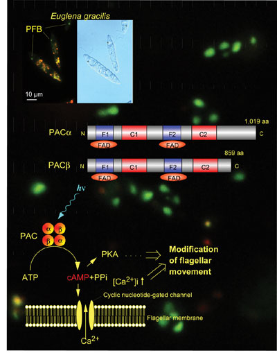
as well as scientific and administrative public services.

(1) Photobiology: Photoreceptive and signal trans-duction mechanisms of phototaxis of unicellular algae are studied action spectroscopically (Watanabe 2004, In “CRC Handbook of Organic Photochemistry and Photobiology, 2nd ed.”) by measuring computerized-videomiceographs of the motile behavior of the cells at the cellular and subcellular levels. Photoreceptive and signal transduction mechanisms of algal gene expression were also studied by action spectroscopy.

A novel blue-light receptor with an effector role was found from Euglena gracilis (Fig. ; Iseki et al. 2002, Nature 415, 1047-1051): Euglena gracilis, a unicellular flagellate, shows blue-light type photomovements. The action spectra indicate the involvement of flavoproteins as the photoreceptors mediating them. The paraflagellar body (PFB), a swelling near the base of the flagellum has been considered as a photosensing organelle for the photomovements. To identify the photoreceptors in the PFB, we isolated PFBs and purified the flavoproteins therein. The purified flavoprotein (ca. 400 kDa), with noncovalently bound FAD, seemed to be a heterotetramer of a- and b-subunits. Predicted amino acid sequences of each of the subunits were similar to each other and contained two FAD-binding domains each followed by an adenylyl cyclase catalytic domain. The flavoprotein showed an adenylyl cyclase activity, which was elevated by blue-light irradiation. Thus, the flavoprotein (PAC, photoactivated adenylyl cyclase) can directly transduce a light signal into a change in the intracellular cyclic AMP level without any other signal transduction proteins.

The involvement of PAC in positive and negative phototaxis (steering response with respect to stimulus light direction) was also demonstrated by its knock-down using RNAi (Ntefidou et al. 2003). Orthologues of PAC a and b were detected in several relatives of Euglena and their phylogenetic analysis indicated that they were trtansfered to euglenoids on the occasion of secondary endosymbiosis (Koumura et al. submitted).

(2) Developmental Biology: Replacement of the ankyrin repeats of mouse Notch2 gene with E. coli b-gactosidase gene induces early embryonic lethality around E10.5. The lethality was suggested due to defects in extraembryonic tissues, because the mutant embryo grew and differentiated further in vitro. Histological examination and in situ hybridization analysis with trophoblast subtype-specific probes revealed that the development of giant and spongiotrophoblast cell layers are normal in the mutant placenta, while vasculogenesis in the labyrinth layer apperaed compromised at E9.5. Since the lethality was circumvented by production of chimeric mice with tetraploidy wild type embryos, we concluded that the embryonic lethality is due to defect in growth and/or differentiation of labyrinthine trophoblast cells. The mutant embryo, however, could not be rescued in the tetraploid chimeras beyond E12.5 because of insurfficient development of umbilical cord, indicating another role of Notch2 signaling in the mouse development. Chimeric analysis with diploid wild type, however, revealed contribution of mutant cells to these affected tissues by E13.5. Thus, Notch2 are not cell autonomously required for the early cell fate determination of labyrinthine trophoblast cells and allantoic mesodermal cells, but plays an indispensable role in the further formation of functional labyrinth layer and umbilical cord.


(3) Computational Biology: Comparative genomics is a useful approach to find clues to understanding complex and diverse biological systems. Our research aim is to develop new methods as well as practical analysis systems to compare a large number of genomic sequences, and to apply them to real data analyses.
Our main developing system is a workbench for microbial genome comparison named MBGD, which now contains more than a hundred of microbial genome sequences. We have continued to enhance the efficiency of the database to treat such large number of genomes. As a key component of MBGD, we have also continued to develop an automated method for orthologous grouping among multiple genomes. In addition to splitting fusion genes into orthologous domains, we are also trying to enhance the algorithm to incorporate some other information such as gene arrangement and species phylogeny.

In addition to these developments, we are also trying to apply the computational methods to real data analyses, especially to the comparative genomics with some closely related organisms. In collabolation with Dr. Takami's group (JAMSTEC), we are comparing genomic sequences of some Bacillus-related organisms that live in several different environmental conditions.

2. Cooperative Research Program for the Okazaki Large Spectrograph

The NIBB Cooperative Research Program for the Use of the OLS supports about 20 projects every year conducted by visiting scientists including foreign scien-tists as well as those in the Institute.
Action spectroscopical studies for various regulatory and damaging actions of light on living organisms, biological molecules, and artificial organic molecules have been conducted (Watanabe, 2004, In “CRC Handbook of Organic Photochemistry and Photobiology, 2nd ed.”. pp. 115-1~115-16).

Publication List:

I. Faculty
Mikami, K. and Murata, N. (2003) Membrane fluidity and the perception of environmental signals in cyanobacteria and plants. Progress Lipid Res. 42, 527-543.

Mikami, K., Suzuki, I. and Murata, N. (2003) Sensors of abiotic stress in Synechocystis. In “Topics in Current Genetics, vol. 4, Plant Stress Response” (H. Hirt and K. Shinozaki eds.), pp 103-119.

Nishiyama T, Fujita T, Shin-I T, Seki M, Nishide H, Uchiyama I, Kamiya A, Carninci P, Hayashizaki Y, Shinozaki K, Kohara Y, Hasebe M. (2003) Comparative genomics of Physcomitrella patens gametophytic transcriptome and Arabidopsis thaliana: implication for land plant evolution. Proc. Natl. Acad. Sci. USA. 100, 8007-8012.

Ntefidou, M., Iseki, M., Watanabe, M., Lebert, M., Haeder, D.-P. (2003) Photoactivated adenylyl cyclase (PAC) controls phototaxis in the flagellate Euglena gracilis. Plant Physiol. 133, 1517-21.

Suzuki, T., Yamasaki, K., Fujita, S., Oda, K., Iseki, M., Yoshida, K., Watanabe, M., Daiyasu, H., Toh, H., Asamizu, E., Tabata, s., Miura, K., Fukuzawa, H., Nakamura, S., Takahashi, T. (2003) Archaeal-type rhodopsins in Chlamydomonas: model structure and intracellular localization. Biochem. Biophys. Res. Commun. 301, 711-717.

Tamura, K., Shimada, T., Ono, Y., Nagatani, A., Higashi, S-I, Watanabe, M., Nishimura, M., Hara-Nishimura, I. (2003) Why green fluorescencent fusion proteins have not been observed in the vacuoles of higher plants. Plant J. 35, 545-555.

Uchiyama, I. (2003) MBGD: microbial genome database for comparative analysis. Nucleic Acids Res. 31, 58-62.

Watanabe, M. (2003) Action spectroscopy for photosensory processes. In “CRC Handbook of Organic Photochemistry and Photobiology, 2nd ed.” (W. Horspool and F. Lenci eds.) pp. 115-1~115-16, CRC Press, Boca Raton

II. Cooperative Research Program for the Okazaki Large Spectrograph
Andrady, A.L., Halis, S. H., and Torikai, A. (2003) Effect of climate change and UV-B on materials. Photochem. Photobiol. Sci. 2, 68-72.

Andrady, A.L., Hamid, S. H., and Torikai, A., (2003) Effect of climate change and UV-B on materials. In “ Environmental Effect of Ozone Depletion and Interactions with Climate Change:2002 Assessment” .

Arimoto-Kobayashi, S., Ando, Y., Horai, Y., Okamoto, K., Hayatsu, H and Michael H. L. G. (2002) Mutation, DNA strand cleavage and nitric oxide formation caused by N-nitroproline with UVA & UVB. J. Photosci. 9, 49-50.

Sasaki, M., Takeshita, S., Oyanagi, T., Miyake, Y. and Sakata, T. (2002). Increasing trend of biologically active solar ultraviolet-B irradiance in mid-latitude Japan in the 1990s. Optical Engineering 41, 3062-3069.

Tamura, K., Shimada, T., Ono, E., Tanaka, Y., Nagatani, A., Higashi, S-i., Watanabe, M., Nishimura, M. and Hara-Nishimura, I. (2003) Why green fluorescent fusion proteins have not been observed in the vacuoles of higher plants. Plant J. 35, 545-555.