NATIONAL INSITUTE FOR BASIC BIOLOGY  


National Institute for Basic Biology

DIVISION OF MOLECULAR NEUROBIOLOGY


Professor:
Masaharu Noda
Associate Professor:
Nobuaki Maeda
Research Associates:
* Masahito Yamagata (~ Sep. 30, 1998)
** Eiji Watanabe (~ Aug. 31, 1998)
Takafumi Shintani (Nov. 1, 1998 ~)
Junichi Yuasa (Nov. 1, 1998 ~)
Post doctral Fellow:
Akihiro Fujikawa 1 (Aug. 16, 1998 ~)
Masakazu Takahashi 4
Hiroyuki Kawachi 4
Hiraki Sakuta 4
Mohamad Zubair 4
Shuhei Kan 4 (Jun. 1, 1998 ~)
Graduate Students:
Chika Saegusa
Akira Kato
Ryoko Suzuki
Visiting Scientists:
*** Elisabeth G. Pollerberg (Mar. 22, 1998 ~ Apr. 19, 1998)
Ikuko Watakabe
Hiroshi Tamura
Technical Staffs:
Akiko Oda (~ Dec. 31, 1998)
Shigemi Takami
JST Technical Staff:
Masae Mizoguchi
Megumi Goto
Minako Ishida (Jan. 1, 1999 ~)
* to Washington University School of Medicine (Oct. 1, 1998 ~)
** to NIBB Center for Transgenic Animals and Plants (Sept. 1, 1998 ~)
*** from Institute for Zoology, University of Heidelberg


We have been studying the molecular and cellular mechanisms underlying the development and functioning of the vertebrate central nervous system. We are currently searching for and analyzing the functions of molecules involved in various cellular events in brain morphogenesis and brain function, such as generation of neuroblasts, their migration to form the laminar structure and various nuclei, elongation and path-finding of neural processes, the formation and refinement of specific connections between neurons, and also synaptic plasticity. We have been using various techniques including molecular biology (e.g. cDNA cloning, site-directed mutagenesis), biochemistry (protein purification, carbohydrate analysis), immunological methods (monoclonal-antibody production), neuroanatomy, cell and organotypic culture (immortalized cell-line production), and embryo manipulation (classical embryology, gene transfer with viral vectors, and gene targeting).


I. Molecular mechanism of retinotectal projection

A. Topographic projection

Topographic maps are a fundamental feature of neural networks in the nervous system. Understanding the molecular mechanisms by which topographically ordered neuronal connections are established during development has long been a major challenge in developmental neurobiology. The retinotectal projection of lower vertebrates including birds has been used as an readily accessible model system. In this projection, the temporal (posterior) retina is connected to the rostral (anterior) part of the contralateral optic tectum, nasal (anterior) retina to the caudal (posterior) tectum, and likewise dorsal and ventral retina are connected to the ventral and dorsal tectum, respectively. Thus, images received by the retina are precisely projected onto the tectum in a reversed manner. In 1963, Sperry proposed that topographic mapping could be guided by complementary position labels in gradients across pre-and postsynaptic fields. Although this concept is widely accepted today, and Eph receptor tyrosine kinases and their ligands were recently identified as candidates for such positional labels, the molecular mechanism of retinotectal map formation remains to be elucidated.

In 1993, we began to screen for topographic molecules which show asymmetrical distribution in the embryonic chicken retina. In the first-round screening, using a cDNA subtractive hybridization technique, we discovered several distinct transcripts which were topographically expressed along the nasotemporal (anteroposterior) axis in the retina. Among these, two winged-helix transcriptional regulators termed CBF-1 and CBF-2 were expressed in the nasal and temporal retina, respectively, and our misexpression experiments using a retroviral vector suggested that these two transcriptional factors direct the retinal ganglion cell axons to project to the appropriate tectal targets along the anteroposterior axis.

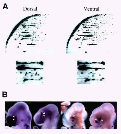
In 1997, to further search for topographic molecules in the embryonic retina, we performed a large-scale screen using a new cDNA display system called Restriction Landmark cDNA Scanning (RLCS) (Fig. 1A). A number of molecules displaying various asymmetrical expression patterns along the nasotemporal axis or dorsoventral axis in the retina have been identified. These included already known topographic molecules such as EphA3, CBF-2, etc. expressed along the nasotemporal axis, and ephrin-B2, EphB3, etc. expressed along the dorsoventral axis.
In 1998, we continued our efforts to identify all of the topographic cDNA clones and examine their expression patterns during development (Fig. 1B). Furthermore, with respect to the topographic molecules which might have important roles in formation of the retinotectal map and/or the neural network in the retina, we have started over- and misexpression experiments using viral vectors and in ovo electroporation.

Fig. 1 Genes expressed in a region-specific fashion in the chick retina
(A) RLCS profiles for the dorsal and ventral retina. The lower panels are enlargements of corresponding regions of the top pictures. Spots indicated by arrows in the left panel are those of dorsal-specific expression. (B) Region-specific gene expression in the retina. Whole-mount in situ hybridization showed that these topographic genes were specifically expressed in the nasal, temporal, dorsal, or ventral region of the retina, respectively.


B. Layer-specific projection

After reaching their appropriate target zones along the rostrocaudal and dorsoventral axes of the tectum, retinal axons begin to seek their appropriate termination sites among 15 distinct laminae within the tectum, of which only three or four receive retinal projections. The molecular and cellular bases of such discrete target choice are poorly understood.

In 1994, we screened for monoclonal antibodies that recognize one of these retinal termination laminae. Among these, we found three clones TB5, TB2 and TB4, which labeled laminae B, D and F, respectively. cDNA cloning and immunochemical analysis revealed that the TB4 antigen molecule was ezrin, a cytoskeletal-membrane linker molecule belonging to the ezrin-radixin-moesin family. Ezrin was selectively expressed in a subset of retinal ganglion cells that project to the lamina F. Similar subset-selective expression and resultant lamina-selective distribution of ezrin were also observed in the lamina-specific central projections from the dorsal root ganglia. The staining pattern for TB4 in the dorsal root ganglia and spinal cord indicated that high expression of ezrin was restricted to cutaneous sensory neurons, but not muscle sensory neurons. Since ezrin modulates cell morphology and cell adhesion profiles by linking specific membrane proteins with the cytoskeleton, it was suggested that ezrin may be involved in the formation and/or maintenance of lamina-specific connections for neuronal subsets in the visual and somatosensory systems.

We expect that our studies will lead to elucidation of the molecular mechanism underlying formation of the retinotectal projection, and ultimately to uncover the basic principles for establishing complicated but extremely precise neural networks in the nervous system.


II. Functional roles of protein tyrosine phosphatase z and g in brain development and brain function

Protein tyrosine phosphorylation plays crucial roles in various aspects of brain development. The level of tyrosine phosphorylation is determined by the balance between the activities of protein tyrosine kinases and protein tyrosine phosphatases. Many types of receptor-like protein tyrosine phosphatases (RPTP) have been cloned and characterized. In 1994, we found that PTPz/RPTPb, a nervous system-specific RPTP, is expressed as a chondroitin sulfate proteoglycan in the brain. An RNA splice variant corresponding to the extracellular region of PTPz is secreted as a major proteoglycan in the brain known as 6B4 proteoglycan/phosphacan. The extracellular region of PTPz consists of a carbonic anhydrase (CAH)-like domain, a fibronectin type III (FN-III)-like domain and a serine-glycine-rich region, which is considered to be the chondroitin sulfate attachment region. PTPz has another family member, RPTPg, which also contains CAH-like and FN-III-like domains. We found that RPTPg has four splice variants including an extracellular secreted form. However, they are not synthesized as proteoglycans and are expressed in various tissues including the brain, kidney, lung and heart.

In 1996, we found that PTPz binds pleiotrophin/heparin-binding growth-associated molecule (HB-GAM) with high affinity (Kd=0.25 nM). The chondroitin sulfate portion of PTPz is essential to achieve high affinity binding between PTPz and pleiotrophin, and removal of chondroitin sulfate chains results in a decrease in the binding affinity (Kd=13 nM). This is the first demonstration that chondroitin sulfate plays an important regulatory role in growth factor signaling.

In the embryonic rat brain, pleiotrophin is localized along the radial glial fibers, a scaffold for neuronal migration. On the other hand, PTPz is expressed in the migrating neurons, suggesting that the ligand-receptor relationship between PTPz and pleiotrophin plays a role in migration of neurons during brain development. Thus, we examined the roles of pleiotrophin-PTPz interaction in neuronal migration using the glass fiber assay and Boyden chamber cell migration assay. Pleiotrophin on the substratum stimulated migration of cortical neurons in both assays. Polyclonal antibodies against the extracellular domain of PTPz, 6B4 proteoglycan (a secreted extracellular form of PTPz) and sodium vanadate (a protein tyrosine phosphatase inhibitor) added to the culture medium strongly suppressed pleiotrophin-induced neuronal migration. These results suggested that PTPz is involved in neuronal migration as a neuronal receptor for pleiotrophin distributed along radial glial fibers.

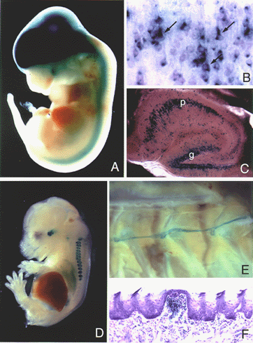
Furthermore, to study the physiological functions of PTPz in vivo, we generated PTPz-deficient mice in which the PTPz gene was replaced by the LacZ gene by homologous recombination in mouse embryonic stem (ES) cells. First, we examined the cell types expressing PTPz by investigating the expression of LacZ in heterozygous PTPz-deficient mice. Throughout development from the early stage of embryogenesis, LacZ staining was restricted to the nervous system. On embryonic day 12.5 (E12.5), LacZ staining was observed in the forebrain, midbrain, hindbrain and spinal cord (Fig. 2A). Examination of the cerebral cortex at higher magnification indicated that subsets of neurons including pyramidal neurons expressed LacZ (Fig. 2B). At the early postnatal stages, subsets of neurons and astrocytes in the brain including pyramidal cells in the hippocampus expressed LacZ (Fig. 2C). Both neurons and astrocytes were positive for LacZ in primary culture of cells from the fetal cerebral cortex. From these results, we concluded that many neurons as well as astrocytes express PTPz. We are currently studying the phenotype of homozygous mutant mice using biochemical, anatomical, physiological and ethological techniques, and various abnormalities have been found.

Fig. 2 LacZ expression in PTPz+/- mice (A-C), and in mNav2.3+/- mice (D-F)
(A) E12.5 whole embryo, (B) P7 cerebral cortex section, and (C) section of adult hippocampus. (D) E15 whole embryo, (E) adult sympathetic nerve and ganglia, and (F) section of adult tongue. In (B), the section was counterstained with cresyl violet, and the arrows indicate the pyramidal cells. In (C), p and g indicate the pyramidal cell layer and granule cell layer, respectively. In (F), the section was counterstained with cresyl violet.


III. Functional roles of subfamily 2 sodium channels

Voltage-gated sodium channels (NaChs) are responsible for the depolarizing phase of action potentials in excitable cells and are essential for many physiological functions. Cloning of NaChs revealed marked conservation in the primary structures that underlies their functional similarity. Thus, all NaChs cloned had been grouped into a single gene family. However, recently, novel NaChs, human Nav2.1, mouse Nav2.3 and rat SCL11/Na-G, were cloned from inexcitable cells such as glial cells. These molecules closely resemble each other but are divergent from the previously cloned sodium channels including the regions involved in activation, inactivation and ion selectivity. Thus, these molecules have been grouped into a new subfamily of sodium channels (subfamily 2). To date, subfamily 2 channels have not been expressed in a functionally active form using in vitro expression systems, and therefore the functional properties of these NaChs are not yet clear. To clarify the cells expressing subfamily 2 sodium channels and their physiological functions in vivo, we planned to generate knock-out mice deficient in channel genes.

W e successfully produced mutant mice in which the mNav2.3 gene was replaced with the LacZ or neo gene by gene targeting. Using these mice, we found that mNav2.3 gene expression was restricted to the dorsal root ganglion (DRG) and lung during the embryonic stage (Fig. 2D). During the postnatal period, in addition to these tissues, Schwann cells in the sensory afferent nerve fibers (Fig. 2E, F) and a subset of neurons in the central nervous system were positive for mNav2.3 expression. We are currently examining the phenotypes of homozygous mNav2.3-deficient mice to gain insight into the physiological functions of this channel.


Publication List:
Shintani, T., Watanabe, E., Maeda, N., and Noda, M. (1998) Neurons as well as astrocytes express proteoglycan-type protein tyrosine phosphatase z/RRTPb: Analysis of mice in which the PTP z/RRTP b gene was replaced with the lacZ gene. Neurosci. Lett. 247, 135-138.
Yamagata, M. and Noda, M. (1998) The winged-helix transcription factor CWH-3 is expressed in developing neural crest cells. Neurosci. Lett. 249, 1-4.
Katoh-Semba, R., Matsuda, M., Watanabe, E., Maeda, N., and Oohira, A. (1998) Two types of brain chondroitin sulfate proteoglycan: their distribution and possible functions in the rat embryo. Neurosci. Res. 31, 273-282.
Maeda, N. and Noda, M. (1998) Involvement of receptor-like protein tyrosine phosphatase z/RPTPb and its ligand pleiotrophin/HB-GAM in neuronal migration. J. Cell Biol., 142, 203-216.
Matsui, F., Nishizuka, M., Yasuda, Y., Aono, S., Watanabe, E. and Oohira, A. (1998) Occurrence of an N-terminal proteolytic fragment of neurocan, not a C-terminal half, in a perineuronal net in the adult rat cerebrum. Brain Res., 790, 45-51.
Nishiwaki, T., Maeda, N. and Noda, M. (1998) Characterization and developmental regulation of proteoglycan-type protein tyrosine phosphatase z/RPTPb isoforms. J. Biochem., 123, 458-467.
Yasuda, Y., Tokita, Y., Aono, S., Matsui, F., Ono, T., Sonta, S., Watanabe, E., Nakanishi, Y., and Oohira, A. (1998) Cloning and chromosomal mapping of the human gene of neuroglycan C (NGC), a neuronal transmembrane chondroitin sulfate proteoglycan with an EGF module. Neurosci. Res., 32, 313-322.
Takahashi, M., Yamagata, M.,and Noda, M. (1999) Specific expression of ezrin, a cytoskeletal-membrane linker protein, in a subset of chick retinotectal and sensory projections. Eur. J. Neurosci. 11, 545-558.
Maeda, N., Ichihara-Tanaka, K., Kimura, T., Kadomatsu, K., Muramatsu, T. and Noda, M. (1999) A receptor-like protein tyrosine phosphatase PTPz/RPTPb binds a heparin-binding growth factor midkine: Involvement of arginine 78 of midkine in the high affinity binding to PTPz. J. Biol. Chem., in press.
Yamakawa, T., Kurosawa, N., Kadomatsu, K., Matsui, T., Itoh, K., Maeda, N., Noda, M., and Muramatsu, T. (1999) Levels of expression of pleiotrophin and protein tyrosine phosphatase z are decreased in human colorectal cancers. Cancer Letter, in press.
Nishiwaki, T., Maeda, N., and Noda, M. (1999) Characterization and developmental regulation of proteoglycan-type protein tyrosine phosphatase z/RPTPb isoforms. In Neural Development (Uyemura, K., Kawamura, K., and Yazaki, T. eds.) pp.291-297. Springer-Verlag Tokyo.


webmaster@nibb.ac.jp
Last Modified: 12:00, May 28, 1999