NATIONAL INSITUTE FOR BASIC BIOLOGY  


National Institute for Basic Biology

DIVISION OF MOLECULAR NEUROBIOLOGY


Professor:
Masaharu Noda
Associate Professor:
Nobuaki Maeda (Oct. 15, 1997 ~)
Research Associates:
Masahito Yamagata
Eiji Watanabe
Post doctral Fellow:
Junichi Yuasa 1
Takafumi Shintani 4
Masakazu Takahashi
Hiroyuki Kawachi 4
Hiraki Sakuta 4 (Oct. 1, 1997 ~)
Graduate Students:
Taeko Nishiwaki
Chika Saegusa
Akira Kato
Ryoko Suzuki (Oct. 1, 1997 ~)
Visiting Scientists:
Angela Mai (Apr. 1 ~ Oct. 31, 1997)
Ikuko Watakabe
Technical Staffs:
Akiko Kawai
Shigemi Ohsugi
JST Technical Staff:
Masae Mizoguchi (May 1, 1997 ~)
Megumi Goto (Jun. 11, 1997 ~)


Our efforts have been devoted to studying molecular and cellular mechanisms underlying the development of the vertebrate central nervous system. We are screening for molecules and structures that regulate various cellular events in brain morphogenesis such as generation of neuroblasts, their migration to form the laminar structure and various nuclei, elongation and path-finding of neural processes, and the formation and refinement of specific connections between neurons. Our research has been conducted using various techniques including molecular biology (e.g. cDNA cloning, site-directed mutagenesis), biochemistry (protein, carbohydrate), immunological methods (monoclonal-antibody production), neuroanatomy, cell and organotypic culture (immortalized cell production), and embryo manipulation (classical embryology, gene transfer with viral vectors, and gene targeting).


I. Topographic map and synapse formation in the retinotectal system

Topographic maps are a fundamental feature of brain organization. In the avian visual system, the temporal (posterior) retina is connected to the rostral (anterior) optic tectum, nasal (anterior) retina to the caudal (posterior) tectum, and likewise dorsal and ventral retina are connected to the ventral and dorsal tectum, respectively. About half a century ago, Sperry proposed that topographic mapping could be guided by complementary positional labels in gradients across pre- and postsynaptic fields. Although this concept is widely accepted today, the developmental mechanism has not been fully characterized at the molecular level. In 1996, using a subtractive hybridization technique, we discovered several distinct transcripts which are topographically expressed in the developing chick retina. Among these molecules, two winged-helix transcriptional regulators termed CBF-1 and CBF-2 were expressed in the nasal and temporal retina, respectively, and our misexpression experiments using a retrovirus vector suggested that these transcription factors direct the retinal ganglion cell axons to choose the appropriate tectal targets along the antero-posterior axis.

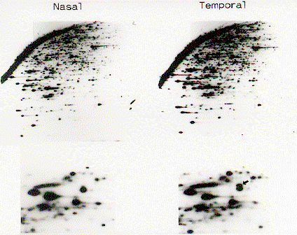
To examine topographic molecules which show asymmetrical distributions in the embryonic retina, we have undertaken a large-scale screen using a new cDNA display system called Restriction Landmark cDNA Scanning (RLCS) (Fig. 1). A number of molecules displaying asymmetrical expression along the naso-temporal axis or dorso-ventral axis in the retina have been identified. This approach seems to be promising because topographic molecules which are already known such as CBF-1, CBF-2, EphA3, aldehyde dehydrogenase etc. were included among those identified. Sequence analyses of the cDNA clones and examination of their expression patterns during development are currently underway. We expect that our studies will lead to systematic identification of a series of topographic molecules in the retina, and to elucidation of the molecular mechanisms underlying formation of the topographic retinotectal projection.

After reaching their appropriate sites along the rostro-caudal and dorso-ventral axes of the tectum, retinal axons begin to seek their appropriate termination sites among 15 distinct laminae within the tectum. The molecular and cellular bases of such discrete target choice are poorly understood, although our previous experiments suggested that molecular complementarity between retinal axons and tectal targets underlies lamina-specific synapse formation. We have generated a series of monoclonal antibodies that recognize one of these retinal termination laminae or a subset of retinal ganglion cells. We hope that characterization of these antigens and their cellular localizations in this relatively accessible system will provide insight into the complicated and still unresolved aspects of neural specificity.

Fig. 1.
RLCS profiles for the chick nasal and temporal retina Topographic molecules were detected as spots specifically found on one side. As an example, see the spot arrowed in the right below panel, the enlargement of a part of the above picture.


II. Receptor-like protein tyrosine phosphatases and brain development

Protein tyrosine phosphorylation plays crucial roles in various aspects of brain development. The level of tyrosine phosphorylation is determined by the balance between the activities of protein tyrosine kinases and protein tyrosine phosphatases. Recently, many types of receptor-like protein tyrosine phosphatases (RPTPs) have been cloned and characterized. In 1994, we found that PTPz/RPTPb, a nervous system-specific RPTP, is expressed as a chondroitin sulfate proteoglycan in the brain. An RNA splice variant corresponding to the extracellular region of PTPz is secreted as a major proteoglycan in the brain known as 6B4 proteoglycan/phosphacan. The extracellular region of PTPz consists of a carbonic anhydrase-like domain, a fibronectin type III-like domain and a serine-glycine-rich region, which is considered to be the chondroitin sulfate attachment region. This unique structure of PTPz has attracted a great deal of attention but little is known about the functional roles of this molecule.

I 1996, we found that PTPz binds pleiotrophin/heparin-binding growth-associated molecule (HB-GAM). In the embryonic rat brain, pleiotrophin is localized along the radial glial fibers, a scaffold for neuronal migration. On the other hand, immunohistochemical studies of normal rat brain indicated that PTPz is expressed in the migrating neurons, suggesting that the ligand-receptor relationship between PTPz and pleiotrophin plays a role in migration of neurons during brain development. Thus, we examined the roles of pleiotrophin-PTPz interaction in neuronal migration using the Boyden chamber cell migration assay. Pleiotrophin coated on the membranes stimulated migration of cortical neurons. Polyclonal antibodies against the extracellular domain of PTPz, 6B4 proteoglycan, a secreted extracellular form of PTPz, and sodium vanadate, a protein tyrosine phosphatase inhibitor, added into the culture medium strongly suppressed the pleiotrophin-induced neuronal migration. Furthermore, chondroitin sulfate C but not chondroitin sulfate A inhibited pleiotrophin-induced neuronal migration, in accordance with our previous findings that chondroitin sulfate constitutes part of the pleiotrophin-binding site of PTPz, and PTPz-pleiotrophin binding is inhibited by chondroitin sulfate C but not by chondroitin sulfate A. These results suggest that PTPz is involved in neuronal migration as a neuronal receptor of pleiotrophin distributed along radial glial fibers.

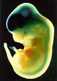
Furthermore, to examine the functions of PTPz in vivo, we generated PTPz-deficient mice in which the PTPz gene was replaced by the LacZ gene by gene targeting. Firstly, we examined the cell types expressing PTPz by investigating the expression of LacZ in heterozygous PTPz-deficient mice. Throughout development from the early stage of embryogenesis, LacZ staining was restricted to the nervous system. At embryonic day 12.5 (E12.5), LacZ staining was observed in the forebrain, midbrain, hindbrain and spinal cord (Fig. 2). In the adult heterozygous mice, LacZ expression was abundant in the olfactory bulb, cerebral cortex, hippocampus, thalamus and cerebellum. In the hippocampus, strong reporter gene expression was observed in the pyramidal neurons and in the granule cell layer of the dentate gyrus. We are currently examining the phenotype of homozygous PTPz-deficient mice.

Fig. 2.
LacZ expression in E12.5 heterozygous PTPz-deficient mice.


III. Gene targeting of Na-G, a voltage-gated sodium channel expressed in glial cells

Glial cells have been considered to be inexcitable. Despite the lack of electrical excitability, they express voltage-gated sodium channels with properties similar to the sodium channels in excitable cells. The cellular function of these voltage-gated sodium channels is not clear. An in vitro study raised the possibility that glial sodium channels serve as a pathway for sodium ion entry to fuel Na-K ATPase, which requires three sodium ions for each pair of potassium ions transported: a drop in the intracellular concentration of sodium ions through this channel may hamper ATPase activity. To test this possibility and clarify the roles of the glial sodium channel in vivo, we are currently attempting to generate a knock-out mouse deficient in the glial sodium channel, Na-G. We have successfully produced chimeric mice which contain the targeted embryonic stem-cells.

By applying gene targeting technology to other novel genes expressed in the brain, we hope to shed light on the molecular mechanisms underlying the development and function of the brain.


Publication List:
Hamanaka, H., Maeda, N. and Noda, M. (1997) Spatially and temporally regulated modification of the receptor-like protein tyrosine phosphatase z/b isoforms with keratan sulfate in the developing chick brain. Eur. J. Neurosci. 9, 2297-2308.
Matsui, F., Nishizuka, M., Yasuda, Y., Aono, S., Watanabe, E. and Oohira,A. (1998) Occurrence of an N-terminal proteolytic fragment of neurocan, not a C-terminal half, in a perineuronal net in the adult rat cerebrum. Brain Res., 790, 45-51 (1998).
Nishiwaki, T., Maeda, N. and Noda, M. (1998) Characterization and developmental regulation of proteoglycan-type protein tyrosine phosphatase z/RPTPb isoforms. J. Biochem., 123, 458-467.
Noda, M., Yamagata, M., Yuasa, J. and Takahashi, M. (1997) Topographic and laminar connection in the chick retinotectal system. In Molecular basis of axon growth and nerve pattern formation (H. Fujisawa, ed.) pp. 197-214. Japan Scientific Societies Press, Tokyo.
Shintani, T., Maeda, N., Nishiwaki, T. and Noda, M. (1997) Characterization of rat receptor-like protein tyrosine phosphatase g isoforms. Biochem. Biophys. Res. Comm. 230, 419-425.


webmaster@nibb.ac.jp
Last Modified: 12:00, May 28, 1998