NATIONAL INSITUTE FOR BASIC BIOLOGY  


National Institute for Basic Biology

DIVISION OF REPRODUCTIVE BIOLOGY


Professor:
Yoshitaka Nagahama
Associate Professor:
Michiyasu Yoshikuni
Research Associates:
Minoru Tanaka
Tohru Kobayashi
Postdoctoral Fellows:
Akihiko Yamaguchi 1
Yoshinao Katsu 2
Mika Tokumoto 2
Takashi Todo 2
Zuxu Yao 2
Craig Morrey 2
JSPS Research Associates:
Daisuke Kobayashi 3
Yuichi Ohba 3
Yasutoshi Yoshiura 3
NIBB Postdoctoral Fellows:
Won-Kyo Lee
Jain-Quiao Jiang
Graduate Students:
Jun Ding (Graduate University for Advanced Studies)
Masatada Watanabe (Graduate University for Advanced Studies)
Gui-jun Guan (Graduate University for Advanced Studies)
Monbusho Foreign Scientist:
Allen Schuetz (Johns Hopkins University)
Visiting Scientist:
Graham Young (University of Otago)


The pituitary-gonadal axis plays an important role in regulating gametogenesis in vertebrates. In most cases, however, gonadotropins act through the biosynthesis of gonadal steroid hormones which in turn mediate various stages of gametogenesis. Studies using teleost fishes as experimental models provide new information about the endocrine regulation of gametogenesis including oocyte growth, oocyte final maturation, spermatogenesis, and sperm maturation. Our research focuses on (1) the identification of steroidal mediators involved in each process of gametogenesis, and (2) the mechanisms of synthesis and action of the mediators. These studies collectively demonstrate the appropriateness of using teleost fishes as valid models for examining hormonal influences on gametogenesis. Such models could also have applications and validity for vertebrates in general.


I. Endocrine regulation of oocyte growth and maturation

17a,20b-dihydroxy-4-pregnen-3-one (17a,20b-DP) was identified as the maturation-inducing hormone of several teleost fishes including salmonid fishes. Along with estradiol-17b, which was identified as the major mediator of oocyte growth, we now have two known biologically important mediators of oocyte growth and maturation in female fishes. Two cell type models have been proposed for estradiol-17b and 17a,20b-DP production, with the thecal layer providing precursor steroids to the granulosa layer. 17a,20b-DP acts via a receptor on the plasma membrane of oocytes. A specific l7a,20b-DP receptor has been identified and characterized from defolliculated oocytes of several fish species. The concentrations of 17a,20b-DP membrane receptors increase immediately prior to oocyte maturation. The pertussis toxin-sensitive G-protein is involved in the signal transduction pathway of 17a,20b-DP. We have cloned two Gia cDNAs from a medaka oocyte cDNA library. Western blot analysis showed that l7a,20b-DP receptor concentrations and Gia content decreased concomitantly in membrane preparations during oocyte maturation. We also found that significant amounts of l7a,20b-DP receptors in the immunoprecipitates, indicating that the l7a,20b-DP membrane receptors are directly coupled with Gi.

The early steps following l7a,20b-DP action involve the formation of the major mediator of this steroid, maturation-promoting factor or metaphase-promoting factor (MPF), which consists of cdc2 kinase (34-kDa) and cyclin B (46- to 48-kDa). Goldfish immature oocytes contain only monomeric 35-kDa inactive cdc2 kinase and do not stockpile cyclin B, although immature oocytes contain mRNA for cyclin B. 17a,20b-DP induces oocytes to synthesize cyclin B, which in turn activates preexisting 35-kDa cdc2 kinase through its threonine 161 phosphorylation by a threonine kinase (MO15), producing the 34-kDa active cdc2. We examined goldfish oocyte proteins bound to cyclin B mRNA. Using oligo(dt)-cellulose affinity chromatograpy and Southwestern and Northwestern analyses, we found a 54-kDa cyclin B mRNA-binding protein (p54), which also bound to the Y box DNA element. We cloned cDNAs encoding Y box proteins, one of which is likely to encode p54 and is expressed specifically in germ cells. We suggest that the 54-kDa Y box protein may have a role in translational repression of cyclin B mRNA in immature oocytes, and 17a,20b-DP stimulation releases this repression, leading to the de nove synthesis of cyclin B.

Immediately prior to the transition from metaphase to anaphase, MPF is inactivated by degradation of cyclin B. We investigated the role of proteasomes (a nonlysosomal large protease) in cyclin degradation, using E. coli-produced goldfish cyclin B and purified goldfish proteasomes (20S and 26S). The purified 26S proteasome, but not 20S proteasome, cleaved both monomeric and cdc2-bound cyclin B at lysine 57 (K57) restrictively in vitro, and produced a 42-kDa N-terminal truncated cyclin B, which was transiently detected at the initial phase of the normal egg activation. The 42-kDa cyclin B, as well as full-length one, was degraded in Xenopus egg extracts, but a mutation on K57 (K57R) inhibited both the digestion by 26S proteasome and the degradation in Xenopus egg extracts. These findings strongly suggest the involvement of 26S proteasome in cyclin degradation through the first cleave on its N-terminus.


II. Endocrine regulation of spermatogenesis

Under cultivation conditions, male Japanese eels (Anguilla japonica) have immature testes containing only type A and early type B spermatogonia together with inactive Sertoli cells and Leydig cells. Using an organ culture system for eel testes, we have shown that the hormonal regulation of spermatogenesis in eel testes involves the gonadotropin stimulation of Leydig cells to produce 11-ketotestosterone (11-KT), a potent androgen in fish. In turn, 11-KT activates Sertoli cells to stimulate premitotic spermatogonia to complete spermatogenesis. Gonadotropin (human chorionic gonadotropin, hCG)-induced spermatogenesis was accompanied by marked, rapid increase in 11-KT production. Northern blot analysis revealed that both 11b-hydroxylase and 11b-hydroxysteroid dehydrogenase mRNA transcripts were not present in testes of eels prior to gonadotropin treatment, but were abundant in those after gonadotropin treatment for 1 day. We used a subtractive hybridization method to clone genes that are expressed differentially in eel testes in the first 24 hr after gonadotropin treatment. One of the up-regulated cDNAs was identified as coding the activin b B subunit. Activn b B mRNA transcripts were absent in testes prior to gonadotropin treatment and were abundant in Sertoli cells in testes of eels treated with gonadotropin for 1-6 days. 11-KT induced a marked production of activin B in cultured testes, indicating that eel activn b B subunit production is largely for activin B formation. Addition of recombinant eel activin B to the culture medium induced proliferation of spermatogonia, producing late type B spermatogonia, within 15 days in the same manner as did 11-KT. Full-length cDNAs for eel activin type I and II receptors were isolated and sequenced. The type I receptor mRNA transcripts were present in testes prior to gonadotropin injection, with a marked increase in spermatogonia and spermatocytes after gonadotropin injection for 9 days. Taken together, these results indicate that activin B produced by Sertoli cells under the influence of gonadotropin and 11-KT plays an important role in the regulation of spermatogenesis in fish testes.

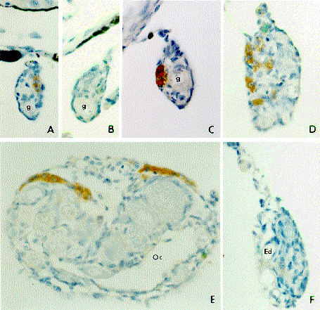
Fig. 1.
Immunocytochemical localization of 3b-hydroxysteroid dehydrogenase and cytochrome P450 aromatase in tilapia gonads collected at 20 days (A, female; B, male), 23-25 days (C, female) , 30 days (D, female), 50 days (E, female), and 30 days (F, male) after hatching. A, B, D, E, and F, 3b-hydroxysteroid dehydrogenase. C, cytochrome P450 aromatase. g, gonium; Oc, ovarian cavity; Ed, efferent duct. X 800.


III. Endocrine regulation of gonadal sex differentiation

The initial differentiation and development of steroid-producing cells during gonadal sexual differentiation in tilapia, Oreochromis niloticus, were examined immunohistochemically using the antibodies of four enzymes essential for steroidogenesis (P450scc, 3b-HSD, P450c17, and P450arom)(Fig. 1). There are two patterns of immunoreactivity in gonads, with either positive reactions for all antibodies, or no reactions with any of the antibodies. Positive immunoreactions for all four antibodies became evident for the first time in the gonads of some fish in mixed sex group examined several days before morphological sex differentiation (about 23 days after hatching). However, positive reactions to the four antibodies increased in number during the process of ovarian differentiation. In contrast, positive immunostaining for all four steroidogenic enzymes could not be confirmed in the testis during sex differentiation. Weakly positive reactions to P450scc, 3b-HSD and P450c17 antibodies appeared first in the testis at 30 days and staining intensity increased slowly during development of the testis. P450arom immunoreactivity was never found. Based on the presence of P450arom, we presume that fish possessing gonads with positive reactions are genetic females and fish with gonads that did not stain with the antibodies are genetic males. These results strongly suggest that endogenous estrogens act as the natural inducers of ovarian differentiation in tilapia. In contrast, the ability of steroid-producing cells to synthesize steroid hormones in the testes appears at the time of testicular differentiation.

Sequential, protogynous hermaphroditism, i.e. female to male sex change in mature fish, exemplifies the plasticity of teleost germ cell differentiation. To begin investigating the importance of hormonal signals on germ cell differentiation during sex change, we examined the expression of P450arom mRNA in the Thalassoma duperrey. Prior to sex change, the gonad of T. duperrey contains primordial germ cells and oogonia but not spermatogonia. P450arom mRNA was detected by Northern analysis and RT-PCR in the ovary. In situ hybridization revealed that P450arom mRNA is localized in the granulosa layer of vitellogenic follicles. These results confirm a major role for aromatase in ovarian physiology. Shortly after the initiation of sex change, however, expression of P450arom mRNA decreases to undetectable levels. P450arom mRNA remains undetectable in the newly-differentiated testis. These results demonstrate that the loss of aromatase is a permanent and critical step in the sex change cascade. As such, they suggest that aromatase/estradiol is important for oogonial differentiation, detrimental to spermatogonial differentiation or a combination of both in T. duperrey.


Publication List:

1. Original Papers
Chang, X.T., Kobayashi, T., Kajiura, H., Nakamura, M. and Nagahama, Y. (1997). Isolation and characterization of the cDNA encoding the tilapia (Oreochromis niloticus) cytochrome P450 aromatase (P450arom): changes in P450arom mRNA, protein and enzyme activity in ovarian follicles during oogenesis. J. Mol. Endocrinol. 18, 57-66.
Ge, W., Peter, R.E. and Nagahama, Y. (1997). Activin and its receptors in the goldfish. Fish Physiol. Biochem. 17, 143-153.
Ge, W., Miura, T., Kobayashi, H., Peter, R.E. and Nagahama, Y. (1997). Cloning of cDNA for goldfish activin b B subunit, and the expression of its mRNA in gonadal and non-gonadal tissues. J. Mol. Endocrinol. 19, 37-45.
Ge, W., Tanaka, M., Yoshikuni, M., Eto, Y. and Nagahama, Y. (1997). Cloning and characterization of goldfish activin type IIB receptor. J. Mol. Endocrinol. 19, 47-57.
Haraguchi, S., Naito, K., Azuma, S., Sato, E., Nagahama, Y., Yamashita, M. and Nagahama, Y. (1996). Effects of phosphate on in vitro 2-cell block of AKR/N mouse embryos based on changes in cdc2 kinase activity and phosphorylation states. Biol. Reprod. 55, 598-603.
Iwao ,Y., Yasumitsu, K., Narihira, M., Jiang, J.-Q. and Nagahama, Y. (1997). Changes in microtubule structures during the first cell cycle of physiologically polyspermic newt eggs. Mol. Reprod. Develop. 47, 210-221.
Katsu, Y., Yamashita, M. and Nagahama, Y. (1997). Isolation and characterization of goldfish Y box protein, a germ-cell-specific RNA binding protein. Eur. J. Biochem. 249, 854-861.
King, P.D., Zhao, Y., Sangoram, A.M., Wilsbacher, L.D., Tanaka, M., Antoch, M.P., Steeves, T.D.L., Vitaterna, M.H., Kornhauser, J.M., Lowrey, P.L., Turek, F.W. and Takahashi, J.S. (1997). Positional cloning of the mouse circadian Clock gene. Cell 89, 641-653.
Matsuyama, M., Morita, S., Hamaji, N., Kashiwagi, M. and Nagahama, Y. (1997). Diurnal spermatogenesis and spawning in the secondary male of a protogynous wrasse, Pseudolabrus japonicus (Teleostie, labridae). Zool Sci. 14, 1001-1008.
Mita. M., Yasumasu, I., Nagahama, Y. and Saneyoshi, M. (1996). Change in the levels of adenine-related compounds in starfish ovarian follicle cells following treatment with gonad-stimulating substance. Dev. Growth Differ. 38, 413-418.
Oba, Y., Yoshikuni, M., Tanaka, M., Mita, M. and Nagahama, Y. (l997). Inhibitory guanine-nucleotide-binding-regulatory protein a subunits in medaka (Oryzias latipes) oocytes: cDNA cloning and decreased expression of proteins during oocyte maturation. Eur. J. Biochem. 249, 846-853.
Tokumoto, M., Horiguchi, R., Yamashita, M., Nagahama, Y. and Tokumoto, T. (1997). Involvement of 26S proteasome in oocyte maturation of goldfish Carassius auratus. Zool. Sci. 14, 347-351.
Tokumoto, T., Yamashita, M., Tokumoto, M., Katsu, Y., Horiguchi, R., Kajiura, H. and Nagahama, Y. (1997). Initiation of cyclin B degradation by the 26S proteasome upon egg activation. J. Cell Biol. 22, 1313-1322.
2. Reveiws and Proceedings
Chang, X.T., Nakamura, M., Kobayashi, T. and Nagahama, Y. (1997). Immunocytochemical localization of steroidogenic enzymes in gonads during sex differentiation in tilapia. J. Reprod. Develop. 43, Suppl. 73-74.
Iwao, Y., Yasumitsu, K., Narihira, M., Jiang, J.Q. and Nagahama, Y. (1997). Changes in microtubule structures in physiologically polyspermic newt eggs. J. Reprod. Develop. 43, Suppl. 157-158.
Mita, M., Yoshikuni, M. and Nagahama, Y. (1997). 1-Methyladenine biosynthesis in ovarian follicle cells of the starfish Asterina pectinifera. In Advances in Comparative Endocrinology (Kawashima, S. and Kikuyama, S., eds.), pp. 317-320, Monduzzi Editore, Bologna, Italy.
Mita, M., Yoshikuni, M. and Nagahama, Y. (1997). Methylation process in 1-methyladenine production by starfish ovarian follicle cells. J. Reprod. Develop. 43, Suppl. 99-100.
Nagahama, Y. (1997). 17a,20b-Dihydroxy-4-pregnen-3-one, a maturation-inducing hormone in fish oocytes: Mechanisms of synthesis and action. Steroids 62, 190-196.
Nagahama, Y., Miura, T., Kobayashi, T. and Ding, J. (1997). The role of activin in spermatogenesis in fish. In Inhibin, Activin and Follistatin (Aono, T., Sugino, H. and Vale, W.W., eds.), pp. 196-203, Springer-Verlag, New York.
Yao, Z., Chang, X.T., Hirai, T., Todo, T., Yoshikuni, M., Kobayashi, T. and Nagahama, Y. (1997). cDNA cloning and protein expression of the gonadotropin receptor from tilapia, Oreochromis niloticus. In Advances in Comparative Endocrinology (Kawashima, S. and Kikuyama, S., eds.), pp. 317-320, Monduzzi Editore, Bologna, Italy.
Young, G., Todo, T., Kobayashi, T., Guan, G. and Nagahama, Y. (1997). Steroidogenesis by the salmonid ovarian follicle: the two-cell type model revisited. In Advances in Comparative Endocrinology (Kawashima, S. and Kikuyama, S., eds.), pp. 1443-1449, Monduzzi Editore, Bologna, Italy.


webmaster@nibb.ac.jp
Last Modified: 12:00, May 28, 1998