NATIONAL INSITUTE FOR BASIC BIOLOGY
![]()
National Institute for Basic Biology
![]() |
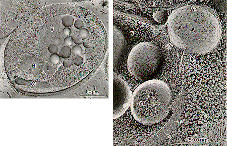
Fig. 1. (a) Freeze-Fracture Image of Yeast under Nitrogen Starvation. When vacuolar proteinases are blocked, spherical membrane structures, autophagic bodies accumulate in the vacuole. (b) Fusion of Autophagosome to the Vacuolar Membrane. Double membrane structure, autophagosome (AP), encloses a portion of the cytosol, then fuses with the vacuole to deliver an autophagic body (AB) in the vacuole. These membrane structures show very few intramembrane particles. |
![]() |
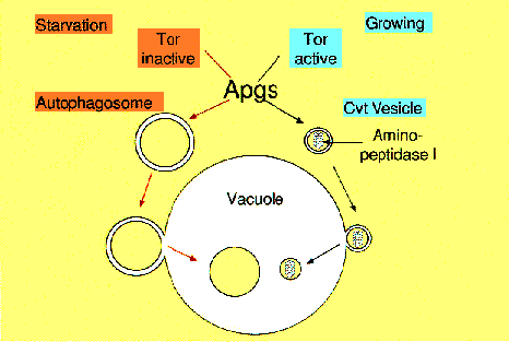
Fig. 2. Schema of autophagy in yeast. When yeast cells face to starvation, isolation membrane starts to enclose a portion of cytosol and form an autophagosome. Autophagosome fuses to the vacuolar membrane, delivering autophagic body in the vacuoles (Autophagic pathway). Under growing condition one of vacuolar enzymes, API is selectively sequestered to the vacuole (Cvt pathway). Two distinct pathways share most molecular machinery. |
webmaster@nibb.ac.jp
Last Modified: 12:00, May 28, 1998