NATIONAL INSITUTE FOR BASIC BIOLOGY  


National Institute for Basic Biology

Division of Gene Expression and Regulation II


Professor:
Takashi Horiuchi
Research Associates:
Masumi Hidaka
Takehiko Kobayashi
Ken-ichi Kodama
Katsuki Johzuka
Graduate Students:
Katufumi Ohsumi
Keiko Taki
Hiroko Urawa
Technical Staffs:
Kohji Hayashi
Yasushi Takeuchi


Homologous recombination which may occur in all organisms apparently involves exchange between two parent-derived chromatids plus the repair of DNA damage incurred by physical and chemical reagents. As deduced from our analyses of recombination hotspots of E. coli and S. cerevisiae, in particular the activity related to DNA replication fork blocking events, the physiological function of homologous recombination, especially in normally growing cells is better understood. In 1996, work on the three following interrelated subjects has advanced our knowledge of related factors.


I. Analysis of E. coli recombinational hot spots

In E. coli RNase H defective (rnh-) mutants, we found specific DNA fragments, termed Hot DNA, when DNA in the ccc form is integrated into the E. coli genome by homologous recombination to form a directly repeated structure and a strikingly enhanced excisional recombination occurs between the repeats. We identified 8 groups (HotA-H) of such Hot DNA, 7 of which (HotA-G) were clustered in a narrow region, known as replication terminus region (about 280 kb) located on the circular E. coli genome. Analysis of HotA, B and C revealed that blocking of replication fork at the Ter, replication terminus, sites is responsible for these Hot activities. Further analysis led to design of a putative model, in which the ds (double stranded)-break occurs at the fork arrested at the DNA replication fork blocking (Ter) site. Through this site the RecBCD recombinational enzyme complex enters the ds-DNA molecule and enhances recombination between directly repeated Hot DNA when the enzyme complex meets an appropriately oriented recombinational hotspot sequence, Chi.

To determine how other termination event-independent Hots can be activated, we chose HotH, because it locates only outside of replication terminus region. HotH is a 11.2 kb EcoRI DNA fragment and locates at about 91 minutes on the E. coli genetic map. We found that it contains the 3' end region of an rRNA operon, rrnD, and also a Chi sequence at the downstream region of rrnD. HotH activity was abolished, when a mutation was introduced into the Chi sequence to destroy its activity, which means that Chi is probably required for HotH activity. We are now investigating events which occur near the HotH site and which probably provide an entrance site for RecBCD enzymes.


II. Atomic structure and functional model of  E. coli replication terminator protein complexed with Ter DNA

Replication of prokaryotic chromosomes is terminated in a defined terminus region. In E. coli, this termination is mediated by a site-specific DNA-binding protein designated as Tus (or tau). The Tus protein (MW 36 kD) is a monomeric molecule that binds to six specific sequences (Ter) within the replication terminus region. The Tus-Ter complex halts passage of replication machinery in only one direction and allows passage in the other direction. This complex controls the passage of DnaB helicase, a constituent of the replication machinery. It is imperative to know the three-dimensional (3D-) structure to gain insight into the mechanism, by which the Tus protein recognizes Ter sites and blocks the replication fork, in a polar manner. Toward this end, we are doing crystallographic studies of the Tus protein in a complex with Ter DNA in collaboration with Drs. K. Kamada and K. Morikawa (Protein Engineering Research Institute, Osaka).

The most suitable crystals for X-ray analysis were grown from a mixture of the Tus protein and a 16 base long DNA duplex, by microdialysis against PEG solution. The crystal structure was determined at 2.7 A resolution by the multiple isomorphous replacement method and anomalous scattering (MIRAS) and has been refined to provide an R-factor of 17.0%.

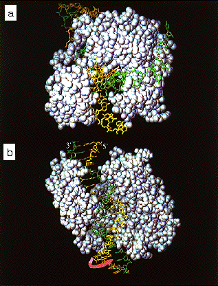
Tus protein adopts an unique structure, as compared with other DNA binding proteins (Figure 1). The structure is divided in two domains, both of which are classified as a + b type. These domains form a positively charged central cleft which mainly accommodates the Ter DNA elements, as if two protruded a-helical regions on both sites and a central b-sheet region would embrace the DNA duplex. The most remarkable feature of the complex is an extensive protein-DNA interface which involves many direct and indirect polar interactions. The two b-sheets of both domains and the interdomain antiparallel b-strands are jointly responsible for recognition of the DNA in the major groove. This structure motif, while unique in detail, is similar to those of MetJ and Arc. The Ter DNA substantially deviates from the canonical B-DNA in the vicinity of central b-bulges of the interdomain b-strands inserted into the major groove. This local deformation of the Ter DNA makes for an overall bend of 20ƒ.

Genetic approaches facilitate the isolation of mutant Tus proteins and most single mutations are mapped to the interdomain region. These mutants that affect efficiency of replication arrest partially or completely impair binding of the Tus protein with DNA. Therefore, the replication arrest and Tus-Ter DNA binding seem to be inseparable.

The overall structural features of the complex favor the interpretation that the a-helical regions of both domains on the fork-blocking side act as a directional DNA clamp and prevent the passage of replication machinery along DNA (Figure 1). Presumably, the passing mechanism from the opposite side relies upon the structural disturbance of the b-sheet region, as induced by the unwinding motion of helicases. This interpretation fits the physical collision model that the intrinsic structure of the complex dominates the polar arrest of the replication fork.

Figure 1.
(a) Tus complexed with a putative Y-forked DNA, as viewed from the fork-block site. The single stranded DNAs near the terminus were hypothetically constructed to realize inhibition of fork progression. (b) A view from the free-passage side. Arrow (pink) denotes the putative unwinding motion of DNA. The unwinding of the helicase from the lower right would displace the yellow DNA strand.


III. The E. coli genome project

E. coli genome project in Japan was started in 1989, the objective being to analyze, in an independently living organism, nucleotide sequences from 0 min in a clockwise direction. In April 1995, the DNA sequences of the 0-12.7 min region in the genetic map were determined and they were published or registered. At that time I (T. H) was appointed leader of the Japan E. coli genome project and a new group of researchers was reorganized to analyze DNA sequences of the region from 12.7 min to 70 min, using Kohara lambda clones and a new protocol, including a shotgun sequence technique.

In January 1997, we determined about a 2.2 mega nucleotide sequence of a region corresponding to 12.7 -69.0 min (including DNA replication terminus region). Combining these data with those (69.0-100 min) provided by Fred Blattner in the USA, the entire E. coli genome was expressed in a single, continuous nucleotide sequence (4,636,552 bp), in which 3803 putative open reading frames were identified. Analysis of the genomic structure by computer is in progress. At this same time, Blattner's group determined independently the entire E. coli DNA sequence. Our sequence data, registered in DNA Data Bank of Japan (DDBJ) are also available through Web sites (http://mol.genes.nig.ac.jp/ecoli/, http://www.ddbj.nig. ac.jp/, http://bsw3.aist-nara.ac.jp/).


Publication List:
Kamada, K., Ohsumi, K., Horiuchi, T., Shimamoto, N. and Morikawa, K. (1996) Crystallization and preliminary X-ray analysis of the Escherichia coli replication terminator protein complexed with DNA. Proteins 24, 402-403.
Ohshima, T., Aiba, H., et al. (1996) A 718-kb DNA sequence of the Escherichia coli K-12 genome corresponding to the 12.7-28.0 min region on the linkage map. DNA Research 3, 1-19.
Kobayashi, T., and Horiuchi, T. (1996) A yeast gene product, Fob1 protein, required for both replication fork blocking and recombinational hotspot activities. Genes to Cells 1, 465-474.
Kamada, K., Horiuchi, T., Ohsumi, K., Shimamoto, N. and Morikawa, K. (1996) Structure of a replication terminator protein complexed with DNA. Nature 383, 598-602.
Aiba, H., Baba, T., et al. (1996) A 570-kb DNA sequence of the Escherichia coli K-12 genome corresponding to the 28.0-40.1 min region on the linkage map. DNA Research 3, 363-377.
Itoh, T., Aiba, H., et al. (1996) A 460-kb DNA sequence of the Escherichia coli K-12 genome corresponding to the 40.1-50.0 min region on the linkage map. DNA Research 3, 379-392.


nibb-adm@nibb.ac.jp
Last Modified: 12:00, June 27, 1997