NATIONAL INSITUTE FOR BASIC BIOLOGY  


National Institute for Basic Biology

Division of Reproductive Biology


Professor:
Yoshitaka Nagahama
Associate Professor:
Michiyasu Yoshikuni
Research Associates:
Minoru Tanaka
Tohru Kobayashi
Institute Research Fellow:
Akihiko Yamaguchi
JSPS Postdoctoral Fellows:
Yoshinao Katsu
Zuxu Yao
NIBB Postdoctoral Fellows:
Kunimasa Suzuki
Won-Kyo Lee
Jain-Quiao Jiang
Xiao-Tian Chang
JSPS Research Associates:
Mika Takahashi
Takashi Todo
Daisuke Kobayashi
Graduate Students:
Yuichi Ohba (Graduate University for Advanced Studies)
Jun Ding (Graduate University for Advanced Studies)
Masatada Watanabe (Graduate University for Advanced Studies)
Guijun Guan (Graduate University for Advanced Studies)
Monbusho Foreign Scientist:
Graham Young (University of Otago)
JSPS Visiting Scientist:
Allen Schuetz (University of Maryland)
Visiting Scientist:
Craig Morrey (University of Hawaii)


The division of reproductive biology conducts research on the endocrine regulation of differentiation, growth and maturation of germ cells in multicellular animals, using fish as a primary study model.


I. Endocrine regulation of oocyte differentiation, growth and maturation

Our research effort in previous years concentrated on the identification and characterization of the molecules (gonadotropins, gonadal steroid hormones, and cell cycle-regulated molecules) that stimulate and control germ cell growth and maturation. We identified, for the first time in any vertebrate, 17a,20b-dihydroxy-4-pregnen-3-one (17a,20b-DP) as the maturation-inducing hormone of amago salmon (Oncorhynchus rhodurus). Along with estradiol-17b, which was identified as the major mediator of oocyte growth, we now have two known biologically important mediators of oocyte growth and maturation in female salmonid fishes. It is established that the granulosa cells are the site of production of these two mediators, but their production by the ovarian follicle depends on the provision of precursor steroids by the thecal cell (two-cell type model). A dramatic switch in the steroidogenic pathway from estradiol-17b to 17a,20b-DP occurs in ovarian follicle cells immediately prior to oocyte maturation. This switch is a prerequisite step for the growing oocyte to enter the maturation phase, and requires a complex and integrated network of gene regulation involving cell-specificity, hormonal regulation, and developmental patterning.

We have isolated and characterized the cDNA encoding several ovarian steroidogenic enzymes of several fish species which are responsible for estradiol-17b and 17a,20b-DP biosynthesis: cholesterol side-chain cleavage cytochrome P450 (P450scc), 3b-hydroxysteroid dehydrogenase (3b-HSD), 17a-hydroxylase/C17,20-lyase cytochrome P450 (P450c17) and P450 aromatase (P450arom). More recently, cDNA clones encoding 20b-hydroxysteroid dehydrogenase (20a-HSD; a critical enzyme which converts 17a-hydroxyprogesterone to 17a,20b-DP) were isolated from cDNA libraries of ayu (Plecoglossus altivelis). The amino acid sequence deduced from the isolated cDNA had 276 amino acid residues and shared approximately 60% homology with mammalian carbonyl reductase. The clear lysate, which was prepared from E. coli harboring the cDNA, catalyzed the production of 17a,20b-DP. The identification of 17a,20b-DP was confirmed by two dimensional thin layer chromatography, followed by recrystallization. Purification of the E. coli-expressed cDNA product revealed that it possessed carbonyl reductase activity and 17a-hydroxyprogesterone, the endogenous immediate precursor of 17a,20b-DP, was a good substrate.

Northern and Western blots revealed a single P450arom mRNA and a single protein in tilapia ovarian tissue respectively. These analyses also revealed that the levels of both P450arom mRNA and protein were low in early vitellogenic follicles, increased in midvitellogenic follicles, and declined to non-detectable levels in postvitellogenic follicles. Changes in the ability of follicles to convert exogenous testosterone to estrogens (aromatase activity) were similar to those of P450arom mRNA and protein. These observations indicated that the capacity of tilapia ovarian follicles to synthesize estradiol-17b is closely related to the contents of P450arom mRNA and protein within them. The mRNA levels of P450scc, 3b-HSD, and P450c17 are barely detected in ovarian follicles during the midvitellogenic stage, and are abundant in follicles during the postvitellogenic stage and oocyte maturation. Our preliminary results indicate that forskolin-induced 17a,20b-DP production is accompanied by a dramatic decrease in P450arom mRNA levels in granulosa cells isolated from postvitellogenic follicles. A 2- to 3-fold increase in P450scc and 3b-HSD mRNAs and a slight decrease in P450c17 mRNA are also observed during forskolin-induced 17a,20b-DP production. Northern hybridization analysis has revealed that 20b-HSD mRNA transcripts are present in fully vitellogenic follicles and increased as oocyte maturation proceeded. Time course studies further suggest that de novo synthesis of 20b-HSD in vitro in response to gonadotropin and cAMP occurs, and consists of gene transcriptional events within the first 6 hr of exposure to gonadotropin and cAMP and translational events 6-9 hr after the exposure to gonadotropin and cAMP. Thus, these results suggest that gonadotropin causes the de novo synthesis of 20b-HSD in the granulosa cell through a mechanism dependent on RNA synthesis.

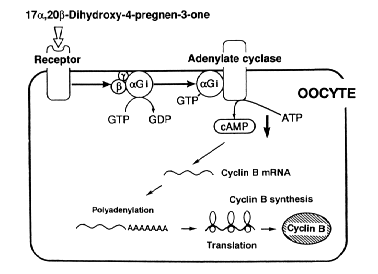
17a,20b-DP acts via a receptor on the plasma membrane of oocytes. We have identified and characterized a specific 17a,20b-DP receptor from defolliculated oocytes of several fish species. Scatchard analysis revealed two different receptors: a high affinity with a Kd of 18 nM and a Bmax of 0.2 pmoles/mg protein and a low affinity receptor with a Kd of 0.5 mM and a Bmax of 1 pmole/mg protein. 17a,20b-DP receptor concentrations increase during oocyte maturation. The interaction between 17a,20b-DP receptors and G-proteins was examined. Pertussis toxin (PT) catalyzed the ADP ribosylation of a 40 kDa protein in crude membranes from rainbow trout oocytes. The 40 kDa protein was recognized by an antibody against the a subunit of inhibitory G-protein. Treating the membrane fraction with 17a,20b-DP decreased the PT-catalyzed ADP ribosylation of the 40 kDa protein. The specific binding of 17a,20b-DP was decreased by PT. We conclude that the PT-sensitive Gi is involved in the signal transduction pathway of 17a,20b-DP in fish oocytes. We have cloned two Gia cDNAs from a medaka oocyte cDNA library. Specific polyclonal antibodies against Gia subunits were generated. These antibodies were used to examine changes in Gia content during 17a,20b-DP-induced oocyte maturation and to immunoprecipitate solubilized Gia. Western blot analysis showed that 17a,20b-DP receptor concentrations and Gia content decreased concomitantly in membrane preparations during oocyte maturation. We also found that significant amounts of 17a,20b-DP receptors in the immunoprecipitates, indicating that the 17a,20b-DP membrane receptors are directly coupled with Gi. This is the first demonstration of direct coupling of the maturation-inducing hormone (steroid) receptor and heteromeric G-proteins (Fig. 1).

Fig. 1
Mechanisms of 17a,20b-dihydroxy-4-pregnen-3-one (17a,20b-DP)-induced cyclin B synthesis by fish oocytes. Inhibitory G-proteins (Gi) is involved in the signal transduction pathway of 17a,20b-DP.


The early steps following 17a,20b-DP action involve the formation of the major mediator of this steroid, maturation-promoting factor or metaphase-promoting factor (MPF). MPF activity cycles during 17a,20b-DP-induced oocyte maturation with the highest activity occurring at the first and second meiotic metaphase. Studies from our laboratory and others have shown that MPF activity is not species-specific and can be detected in both meiotic and mitotic cells of various organisms, from yeast to mammals.

Fish MPF, like that of amphibians, consists of two components, catalytic cdc2 kinase (34-kDa) and regulatory cyclin B (46- to 48-kDa). Goldfish immature oocytes contain 35-kDa inactive cdc2 kinase. Although immature oocytes contain mRNA for cyclin B, they do not contain cyclin B protein. 17a,20b-DP induces oocytes to synthesize cyclin B. The preexisting 35-kDa inactive cdc2 kinase binds to de novo synthesized cyclin B at first, then is rapidly converted into the 34-kDa active form. Introduction of a bacterially produced goldfish cyclin B into immature goldfish oocyte extracts induces cdc2 kinase activation. Phosphoaminoacid analysis shows that threonine (Thr) phosphorylation of the 34-kDa cdc2 kinase is associated with the activation. The sites of Thr phosphorylation on cdc2 kinase was mapped to reside Thr-161. Since goldfish cyclin B mRNA contains four copies of the usual cytoplasmic polyadenylation element in the 3'UTR, the initiation of its synthesis during oocyte maturation may be controlled by the elongation of poly (A) tail. We examined the polyadenylation state of cyclin B mRNA during goldfish oocyte maturation by means of a PCR poly (A) test, and found that cyclin B mRNA is polyadenylated during oocyte matuation. Furthermore, cordycepin which inhibits poly (A) addition of mRNA, prevented 17a,20b-DP-induced oocyte maturation in goldfish. It is concluded that elongation of poly (A) tail of cyclin B mRNA is required for the initiation of its translation (Fig. 1).

Immediately prior to the transition from metaphase to anaphase, MPF is inactivated by degradation of cyclin B. We investigated the role of proteasomes (a nonlysosomal large protease) in cyclin degradation, using E. coli-produced goldfish cyclin B and purified goldfish proteasomes (20S and 26S). The purified 26S proteasome, but not 20S proteasome, cleaved both monomeric and cdc2-bound cyclin B at lysine 57 (K57) restrictively in vitro, and produced a 42 kDa N-terminal truncated cyclin B, which was transiently detected at the initial phase of the normal egg activation. The 42 kDa cyclin B, as well as full-length one, was degraded in Xenopus egg extracts, but a mutation on K57 (K57R) inhibited both the digestion by 26S proteasome and the degradation in Xenopus egg extracts. These findings strongly suggest that the involvement of 26S proteasome in cyclin degradation through the first cleave on its N-terminus.


II. Endocrine regulation of male germ cell development and maturation

We have identified two steroidal mediators of male germ cell development in salmonid fishes (11-ketotestosterone for spermatogenesis and 17a,20b-DP for sperm maturation). A steroidogenic switch, from 11-ketotestosterone to 17a,20b-DP, occurs in salmonid testes around the onset of final maturation. In vitro incubation studies using different testicular preparations have revealed that the site of 17a,20b-DP production is in the sperm, but its production depends on the provision of precursor steroids by somatic cells. The site of 11-ketotestosterone production is in the testicular somatic cells.

In the cultivated male Japanese eel (Auguilla japonica), spermatogonia are the only germ cells present in the testis. A serum-free, chemically defined organ culture system developed for eel testes was used to investigate the effect of various steroid hormones on the induction of spermatogenesis in vitro. We obtained evidence that 11-ketotestosterone can induce the entire process of spermatogenesis in vitro from premitotic spermatogonia to spermatozoa within 21 days.

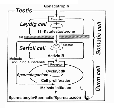
We have used subtractive hybridization to identify genes that are expressed differentially in eel testes in the first 24 hr after HCG treatment in vivo, which ultimately induces spermatogenesis. One up-regulated cDNA was isolated from subtractive cDNA libraries derived from mRNA extracted from control testes and testes one day after a single injection of HCG. From its deduced amino acid sequence, this clone was identified as coding for the activin bB subunit. Using Northern blot analysis and in situ hybridization techniques, we examined sequential changes in transcripts of testicular activin bB during HCG-induced spermatogenesis. No transcripts for activin bB were found in testes prior to HCG injection. In contrast, 3.3 kb mRNA transcripts were prominent in testes one day after the injection. The transcript concentration began to decrease three days after the injection and there was a further sharp decrease by nine days. The HCG-dependent activin bB mRNA expression in the testes was confirmed by in situ hybridization using a digoxigenin-labelled RNA probe: the signal was restricted to Sertoli cells in testes treated with HCG for one to three days. A marked stimulation of activin B production, but not either activin A or activin AB, was observed in testes after HCG and 11-ketotestosterone treatment. Addition of recombinant human and eel activin B to the culture medium induced proliferation of spermatogonia, producing mitotic spermatogonia, within 15 days in the same manner as did 11-ketotestosterone. Taken together, these findings suggest the following sequence of the hormonal induction of spermatogenesis in the eel. Gonadotropin stimulates the Leydig cells to produce 11-ketotestosterone, which, in turn, activates the Sertoli cells to produce activin B. Activin B then acts on spermatogonia to induce mitosis leading to the formation of spermatocytes (Fig. 2).

Fig. 2.
Hormonal regulation of spermatogenesis in the eel testis.


To characterize the pathway of activin signal transduction involved in the process of spermatogenesis, we recently initiated studies on the cloning and expression of activin receptors of eel testes. Three kinds of cDNA clones encoding eel activin type I receptors were isolated from an eel testis cDNA library. The predicted proteins of these receptors consist of 510, 493, and 505 amino acids with their deduced amino acid sequences having a putative signal peptide, a cystein-rich extracellular domain containing a potential N-glycosylation site, a putative transmembrane domain with a well conserved GSGSG motif, and a serine/threonine kinase domain. Northern blot hybridization was performed in testes from eels injected with hCG for 1-18 days. The results were that mRNA transcripts of the three eel activin type I receptors were expressed in the testes of eels prior to hCG injection, with marked increased in the testes after hCG injection for 9 days. In situ hybridization revealed that these activin type I receptors are localized in germ cells (spermatogonia and spermatocytes) and Sertoli cells. To further understand the molecular mechanisms of activin B-induced initiation of mitosis in spermatogonia, we recently isolated cDNA clones encoding eel homologs of cdc2 kinase, cdk2 kinase, cdk4 kinase, cyclin A1 and A2, cyclin B1 and B2, cyclin D1, and cyclin E and E' from cDNA libraries of eel testes. Specific antibodies against some of these proteins were also raised.


III. Endocrine regulation of gonadal sex differentiation

Recently, we, in collaboration with Drs. K. Ozato (Nagoya University), M. Nakamura (Teikyo University) and E.G. Grau (University of Hawaii), have initiated studies on the roles of sex steroid hormones in sex determination and differentiation using medaka, tilapia (Oreochromis niloticus), and a sex change fish, Thalassoma duperrey. In tilapia, a pair of gonadal primordia formed on both sides of the intestine at 15 days after hatching. The gonadal anlage consists of several roundish germ cells surrounded by a few stromal cells. At 20 days after hatching, prior to the period of sex differentiation, positive immunostaining for P450scc, 3b-HSD, P450c17, and P450arom antibodies becomes evident for the first time in gonads. Immunostained stromal cells are observed in the vicinity of blood vessels. From 23-26 days after hatching, morphological gonadal sex differentiation begins to be recognized. Initial ovarian differentiation is marked by the appearance of a narrow space in the stromal tissue, representing the formation of the ovarian cavity. On the other hand, initial testicular differentiation is characterized by the appearance of a narrow space in the stromal tissue, representing the efferent duct construction. At this stage, germ cells in testes and ovaries remain in the gonial stage. In ovaries, positive immunostaining for four kinds of steroidogenic enzymes is recognized in large stromal cells located in the vicinity of blood vessels. In contrast, no immunoreaction is evident in testes during sex differentiation. This situation continues until testes initiate spermatogenesis. Thus, tilapia ovaries express the steroidogenic enzymes required for estradiol-17b biosynthesis from cholesterol before sexual differentiation, which is consistent with the concept that estrogen biosynthesis is essential for sexual differentiation of female phenotype during early development.

In T. duperrey, steroid-producing cells have identified at different stages of sex change using immunocytochemical methods. Immunoreactive P450scc, the first enzyme in the steroidogenic pathway, initially localizes in thecal cells. As sex change progresses towards testis formation, P450scc becomes localized in interstitial cells. It appears that thecal cells migrate towards the interstices and become very active androgen-producing cells.


Publication List:
Chang, X.-T., Kobayashi, T., Kajiura, H., Nakamura, M. and Nagahama, Y. (1996). Isolation and characterization of the cDNA encoding the tilapia (Oreochromis niloticus) cytochrome P450 aromatase (P450arom): changes in P450arom mRNA, protein and enzyme activity in ovarian follicle during oogenesis. J. Mol. Endocrinol. 18, 57-66.
Fukada, S., Tanaka, M., Matsuyama, M., Kobayashi, D. and Nagahama, Y. (1996). Isolation, characterization, and expression of cDNAs encoding the medaka (Oryzias latipes) ovarian follicle cytochrome P-450 aromatase. Mol. Reprod. Develop. 45, 285-290.
Jiang, J.-Q., Kobayashi, T., Ge, W., Kobayashi, H., Tanaka, M., Okamoto, M., Nonaka, Y. and Nagahama, Y. (1996). Fish testicular 11b-hydroxylase: cDNA cloning and mRNA expression during spermatogenesis. FEBS Letters 397, 250-252.
Jiang, J.-Q., Yamashita, M., Yoshikuni, M., Fukada, S. and Nagahama, Y. (1996). Organization of cytoplasmic microtubules during maturation of goldfish oocytes. Zool. Sci. 13, 899-907.
Kobayashi, T., Chang, X.-T., Nakamura, M., Kajiura, H. and Nagahama, Y. (1996). Fish 3b-hydroxysteroid dehydrogenase/D5-D4isomerase: antibody production and their use for the immunohistochemical detection of fish steroidogenic tissues. Zool. Sci. 13, 909-914.
Kobayashi, D., Tanaka, M., Fukada, S. and Nagahama, Y. (1996). Steroidogenesis in the ovarian follicles of the medaka (Oryzias latipes) during vitellogenesis and oocyte maturation. Zool. Sci. 13, 921-927.
Mineyuki, Y., Aioi, H., Yamashita, M. and Nagahama, Y. (1996). A comparative study on stainability of preprophase bands by the PSTAIR antibody. J. Plant Res. 109, 185-192.
Miura, C., Miura, T., Yamauchi, K. and Nagahama, Y. (l996). Hormonal induction of all stages of spermatogenesis in germ-somatic cell cultures from immature Japanese eel testis. Develop. Growth Differ. 38, 257-262.
Nagahama, Y., Miura, T., Kobayashi, T. and Ding J. (1996). The role of activin in spermatogenesis in fish. Proceedings of Serono Symposium (in press).
Nagahama, Y. (1996). 17a,20b-Dihydroxy-4-pregnen-3-one, a maturation-inducing hormone in fish oocytes: Mechanisms of synthesis and action. Steroid 62, 190-196.
Nakamura, M., Specker, J.L. and Nagahama, Y. (1996). Innervation of steroid-producing cells in the ovary of tilapia Oreochromis niloticus. Zool. Sci. 13, 603-608.
Nogami, A., Suzaki, T., Shigenaka, Y., Nagahama, Y. and Mineyuki, Y. (1996). Effects of cycloheximide on preprophase bands and prophase spindles in onion (Allium cepa L.) root tip cells. Protoplasma 192, 109-121.
Sakai, N., Suzuki, N. and Nagahama Y. (1996). Distribution of the site of 17a,20b-dihydroxy-4-pregnen-3-one production in the spermiating amago salmon (Oncorhynchus rhodurus) testis. Biomed. Res. 17, 473-477.
Sakai, N., Suzuki, N. and Nagahama, Y. (1996). Involvement of cyclic AMP in the regulation of testicular steroidogenesis of amago salmon (Oncorhynchus rhodurus). Biomed. Res. 17, 479-486.


nibb-adm@nibb.ac.jp
Last Modified: 12:00, June 27, 1997